2RKU | pdb_00002rku

Structure of PLK1 in complex with BI2536


Experimental Data Snapshot

  • Method: X-RAY DIFFRACTION
  • Resolution: 1.95 Å
  • R-Value Free: 
    0.241 (Depositor), 0.243 (DCC) 
  • R-Value Work: 
    0.190 (Depositor), 0.205 (DCC) 
  • R-Value Observed: 
    0.192 (Depositor) 

wwPDB Validation 3D Report Full Report

Validation slider image for 2RKU

Ligand Structure Quality Assessment 


This is version 1.4 of the entry. See complete history

Literature

Selectivity-determining residues in Plk1.

Kothe, M.Kohls, D.Low, S.Coli, R.Rennie, G.R.Feru, F.Kuhn, C.Ding, Y.-H.

(2007) Chem Biol Drug Des 70: 540-546

  • DOI: https://doi.org/10.1111/j.1747-0285.2007.00594.x
  • Primary Citation Related Structures: 
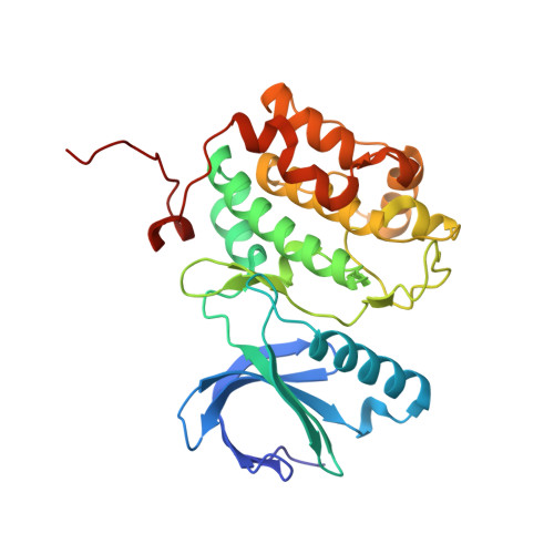
    2RKU

  • PubMed Abstract: 

    Polo-like kinase 1 is an important regulator of cell cycle progression whose over-expression is often associated with oncogenesis. Polo-like kinase 1 hence represents an attractive target for cancer intervention. BI 2536 (Boehringer Ingelheim, Ingelheim, Germany), a Polo-like kinase 1 inhibitor currently in clinical trials, exhibits nanomolar potency against Polo-like kinase isoforms and high selectivity against other kinases. We have previously published the crystal structures of the Polo-like kinase 1 domain in complex with AMPPNP and an Aurora A inhibitor. In this work, we present the co-crystal structure of Polo-like kinase 1 with BI 2536. The structure, in combination with selectivity data for BI 2536 and related compounds, illustrates important features for potency and selectivity. In particular, we show that the methoxy group of BI 2536 is an important specificity determinant against non-Polo-like kinases by taking advantage of a small pocket generated by Leu 132 in the hinge region of Polo-like kinase 1. The work presented here provides a framework for structure-based drug design of Polo-like kinase 1-specific inhibitors.


  • Organizational Affiliation
    • Pfizer Global Research and Development, Research Technology Center, 620 Memorial Drive, Cambridge, MA 02139, USA.

Macromolecule Content 

  • Total Structure Weight: 35.16 kDa 
  • Atom Count: 2,785 
  • Modeled Residue Count: 294 
  • Deposited Residue Count: 294 
  • Unique protein chains: 1

Macromolecules

Find similar proteins by:|  3D Structure
Entity ID: 1
MoleculeChains  Sequence LengthOrganismDetailsImage
Serine/threonine-protein kinase PLK1294Homo sapiensMutation(s): 1 
Gene Names: PLK1PLK
EC: 2.7.11.21
UniProt & NIH Common Fund Data Resources
Find proteins for P53350 (Homo sapiens)
Explore P53350 
Go to UniProtKB:  P53350
PHAROS:  P53350
GTEx:  ENSG00000166851 
Entity Groups
Sequence Clusters30% Identity50% Identity70% Identity90% Identity95% Identity100% Identity
UniProt GroupP53350
Sequence Annotations
Expand
Reference Sequence

Small Molecules

Ligands 5 Unique
IDChains Name / Formula / InChI Key2D Diagram3D Interactions
R78

Query on R78


Download:Ideal Coordinates CCD File
C [auth A]4-{[(7R)-8-cyclopentyl-7-ethyl-5-methyl-6-oxo-5,6,7,8-tetrahydropteridin-2-yl]amino}-3-methoxy-N-(1-methylpiperidin-4-yl)benzamide
C28 H39 N7 O3
XQVVPGYIWAGRNI-JOCHJYFZSA-N
TAR

Query on TAR


Download:Ideal Coordinates CCD File
H [auth A]D(-)-TARTARIC ACID
C4 H6 O6
FEWJPZIEWOKRBE-LWMBPPNESA-N
SRT

Query on SRT


Download:Ideal Coordinates CCD File
F [auth A],
G [auth A]
S,R MESO-TARTARIC ACID
C4 H6 O6
FEWJPZIEWOKRBE-XIXRPRMCSA-N
TLA

Query on TLA


Download:Ideal Coordinates CCD File
D [auth A],
E [auth A]
L(+)-TARTARIC ACID
C4 H6 O6
FEWJPZIEWOKRBE-JCYAYHJZSA-N
ZN

Query on ZN


Download:Ideal Coordinates CCD File
B [auth A]ZINC ION
Zn
PTFCDOFLOPIGGS-UHFFFAOYSA-N

Experimental Data & Validation

Experimental Data

  • Method: X-RAY DIFFRACTION
  • Resolution: 1.95 Å
  • R-Value Free:  0.241 (Depositor), 0.243 (DCC) 
  • R-Value Work:  0.190 (Depositor), 0.205 (DCC) 
  • R-Value Observed: 0.192 (Depositor) 
Space Group: P 32 2 1
Unit Cell:
Length ( Å )Angle ( ˚ )
a = 66.593α = 90
b = 66.593β = 90
c = 154.11γ = 120
Software Package:
Software NamePurpose
DENZOdata reduction
SCALEPACKdata scaling
REFMACrefinement
PDB_EXTRACTdata extraction
HKL-2000data reduction
HKL-2000data scaling

Structure Validation

View Full Validation Report


Ligand Structure Quality Assessment 


Entry History 

Deposition Data

Revision History  (Full details and data files)

  • Version 1.0: 2008-02-05
    Type: Initial release
  • Version 1.1: 2011-07-13
    Changes: Advisory, Version format compliance
  • Version 1.2: 2017-10-25
    Changes: Refinement description
  • Version 1.3: 2021-10-20
    Changes: Database references, Derived calculations
  • Version 1.4: 2024-02-21
    Changes: Data collection