


Crystal structure of dipicolinate synthase, A chain, from Bacillus subtilis.
Osipiuk, J., Quartey, P., Moy, S., Joachimiak, A.To be published.
Experimental Data Snapshot
Macromolecule Content
Entity ID: 1 | |||||
|---|---|---|---|---|---|
| Molecule | Chains | Sequence Length | Organism | Details | Image |
| Dipicolinate synthase, A chain | 300 | Bacillus subtilis | Mutation(s): 0 Gene Names: spoVFA, dpaA, BSU16730 EC: 1.3.1 | 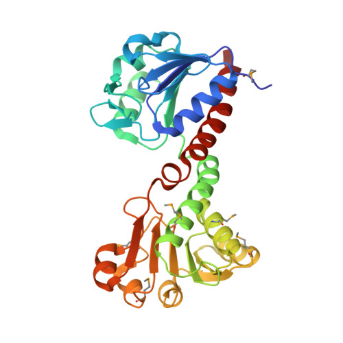 | |
UniProt | |||||
Entity Groups | |||||
| Sequence Clusters | 30% Identity50% Identity70% Identity90% Identity95% Identity100% Identity | ||||
| UniProt Group | Q04809 | ||||
Sequence AnnotationsExpand | |||||
Reference Sequence | |||||
| Ligands 2 Unique | |||||
|---|---|---|---|---|---|
| ID | Chains | Name / Formula / InChI Key | 2D Diagram | 3D Interactions | |
| NAP Download:Ideal Coordinates CCD File | J [auth A] L [auth B] M [auth C] O [auth D] Q [auth E] | NADP NICOTINAMIDE-ADENINE-DINUCLEOTIDE PHOSPHATE C21 H28 N7 O17 P3 XJLXINKUBYWONI-NNYOXOHSSA-N |  | ||
| CL Download:Ideal Coordinates CCD File | I [auth A] K [auth B] N [auth D] P [auth E] S [auth G] | CHLORIDE ION Cl VEXZGXHMUGYJMC-UHFFFAOYSA-M |  | ||
| Modified Residues 1 Unique | |||||
|---|---|---|---|---|---|
| ID | Chains | Type | Formula | 2D Diagram | Parent |
| MSE Query on MSE | A, B, C, D, E A, B, C, D, E, F, G, H | L-PEPTIDE LINKING | C5 H11 N O2 Se |  | MET |
| Length ( Å ) | Angle ( ˚ ) |
|---|---|
| a = 261.578 | α = 90 |
| b = 75.108 | β = 119.74 |
| c = 200.464 | γ = 90 |
| Software Name | Purpose |
|---|---|
| DENZO | data reduction |
| SCALEPACK | data scaling |
| REFMAC | refinement |
| PDB_EXTRACT | data extraction |
| SBC-Collect | data collection |
| HKL-2000 | data reduction |
| SHELXD | phasing |
| SOLVE | phasing |
| RESOLVE | phasing |
| HKL-2000 | data scaling |


