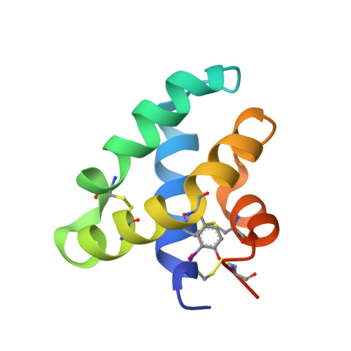
Crystal structures of human saposins C and d: implications for lipid recognition and membrane interactions.
Rossmann, M., Schultz-Heienbrok, R., Behlke, J., Remmel, N., Alings, C., Sandhoff, K., Saenger, W., Maier, T.(2008) Structure 16: 809-817
- PubMed: 18462685
- DOI: https://doi.org/10.1016/j.str.2008.02.016
- Primary Citation of Related Structures:
2QYP, 2R0R, 2R1Q, 2RB3, 2Z9A - PubMed Abstract:
Human saposins are essential proteins required for degradation of sphingolipids and lipid antigen presentation. Despite the conserved structural organization of saposins, their distinct modes of interaction with biological membranes are not fully understood. We describe two crystal structures of human saposin C in an "open" configuration with unusual domain swapped homodimers. This form of SapC dimer supports the "clip-on" model for SapC-induced vesicle fusion. In addition, we present the crystal structure of SapD in two crystal forms. They reveal the monomer-monomer interface for the SapD dimer, which was confirmed in solution by analytical ultracentrifugation. The crystal structure of SapD suggests that side chains of Lys10 and Arg17 are involved in initial association with the preferred anionic biological membranes by forming salt bridges with sulfate or phosphate lipid headgroups.
- Institut für Chemie und Biochemie, Kristallographie, Freie Universität Berlin, Takustr. 6, 14195 Berlin, Germany.
Organizational Affiliation:


