2QVA

Crystal structure of Drosophila melanogaster Translin protein


Experimental Data Snapshot

  • Method: X-RAY DIFFRACTION
  • Resolution: 3.40 Å
  • R-Value Free: 0.269 
  • R-Value Work: 0.203 
  • R-Value Observed: 0.207 

wwPDB Validation   3D Report Full Report


This is version 1.3 of the entry. See complete history


Literature

Crystal structures of Drosophila mutant translin and characterization of translin variants reveal the structural plasticity of translin proteins.

Gupta, G.D.Makde, R.D.Rao, B.J.Kumar, V.

(2008) FEBS J 275: 4235-4249

  • DOI: https://doi.org/10.1111/j.1742-4658.2008.06571.x
  • Primary Citation of Related Structures:  
    2QRX, 2QVA

  • PubMed Abstract: 

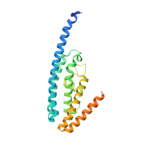
    Translin protein is highly conserved in eukaryotes. Human translin binds both ssDNA and RNA. Its nucleic acid binding site results from a combination of basic regions in the octameric structure. We report here the first biochemical characterization of wild-type Drosophila melanogaster (drosophila) translin and a chimeric translin, and present 3.5 A resolution crystal structures of drosophila P168S mutant translin from two crystal forms. The wild-type drosophila translin most likely exists as an octamer/decamer, and binds to the ssDNA Bcl-CL1 sequence. In contrast, ssDNA binding-incompetent drosophila P168S mutant translin exists as a tetramer. The structures of the mutant translin are identical in both crystal forms, and their C-terminal residues are disordered. The chimeric protein, possessing two nucleic acid binding motifs of drosophila translin, the C-terminal residues of human translin, and serine at position 168, attains the octameric state and binds to ssDNA. The present studies suggest that the oligomeric status of translin critically influences the DNA binding properties of translin proteins.


  • Organizational Affiliation

    High Pressure Physics Division, Bhabha Atomic Research Centre, Mumbai, India.


Macromolecules
Find similar proteins by:  (by identity cutoff)  |  3D Structure
Entity ID: 1
MoleculeChains Sequence LengthOrganismDetailsImage
GM27569p
A, B, C, D
247Drosophila melanogasterMutation(s): 1 
Gene Names: translin
UniProt
Find proteins for Q7JVK6 (Drosophila melanogaster)
Explore Q7JVK6 
Go to UniProtKB:  Q7JVK6
Entity Groups  
Sequence Clusters30% Identity50% Identity70% Identity90% Identity95% Identity100% Identity
UniProt GroupQ7JVK6
Sequence Annotations
Expand
  • Reference Sequence
Experimental Data & Validation

Experimental Data

  • Method: X-RAY DIFFRACTION
  • Resolution: 3.40 Å
  • R-Value Free: 0.269 
  • R-Value Work: 0.203 
  • R-Value Observed: 0.207 
  • Space Group: P 2 2 21
Unit Cell:
Length ( Å )Angle ( ˚ )
a = 98.58α = 90
b = 96.62β = 90
c = 153.41γ = 90
Software Package:
Software NamePurpose
REFMACrefinement
MAR345dtbdata collection
MOSFLMdata reduction
SCALAdata scaling
AMoREphasing

Structure Validation

View Full Validation Report


Entry History 

Deposition Data

Revision History  (Full details and data files)

  • Version 1.0: 2008-08-05
    Type: Initial release
  • Version 1.1: 2011-07-13
    Changes: Version format compliance
  • Version 1.2: 2021-11-10
    Changes: Database references
  • Version 1.3: 2023-10-25
    Changes: Data collection, Refinement description