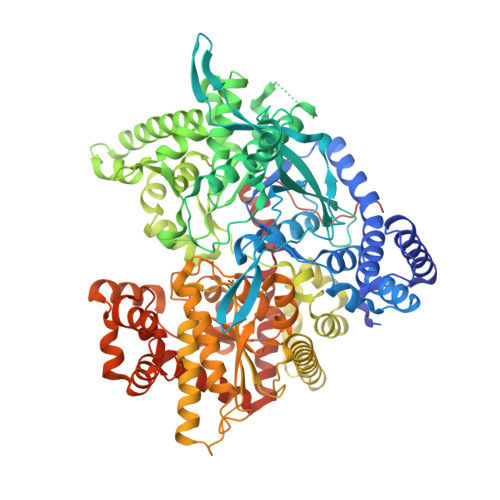
2QRG

Glycogen Phosphorylase b in complex with (1R)-3'-(4-methoxyphenyl)-spiro[1,5-anhydro-D-glucitol-1,5'-isoxazoline]


Experimental Data Snapshot

  • Method: X-RAY DIFFRACTION
  • Resolution: 1.85 Å
  • R-Value Free: 0.211 
  • R-Value Work: 0.190 
  • R-Value Observed: 0.191 

Starting Model: experimental
View more details

wwPDB Validation   3D Report Full Report


This is version 1.3 of the entry. See complete history


Literature

Glucose-based spiro-isoxazolines: a new family of potent glycogen phosphorylase inhibitors.

Benltifa, M.Hayes, J.M.Vidal, S.Gueyrard, D.Goekjian, P.G.Praly, J.P.Kizilis, G.Tiraidis, C.Alexacou, K.M.Chrysina, E.D.Zographos, S.E.Leonidas, D.D.Archontis, G.Oikonomakos, N.G.

(2009) Bioorg Med Chem 17: 7368-7380

  • DOI: https://doi.org/10.1016/j.bmc.2009.08.060
  • Primary Citation of Related Structures:  
    2QRG, 2QRH, 2QRM, 2QRP, 2QRQ

  • PubMed Abstract: 

    A series of glucopyranosylidene-spiro-isoxazolines was prepared through regio- and stereoselective [3+2]-cycloaddition between the methylene acetylated exo-glucal and aromatic nitrile oxides. The deprotected cycloadducts were evaluated as inhibitors of muscle glycogen phosphorylase b. The carbohydrate-based family of five inhibitors displays K(i) values ranging from 0.63 to 92.5 microM. The X-ray structures of the enzyme-ligand complexes show that the inhibitors bind preferentially at the catalytic site of the enzyme retaining the less active T-state conformation. Docking calculations with GLIDE in extra-precision (XP) mode yielded excellent agreement with experiment, as judged by comparison of the predicted binding modes of the five ligands with the crystallographic conformations and the good correlation between the docking scores and the experimental free binding energies. Use of docking constraints on the well-defined positions of the glucopyranose moiety in the catalytic site and redocking of GLIDE-XP poses using electrostatic potential fit-determined ligand partial charges in quantum polarized ligand docking (QPLD) produced the best results in this regard.


  • Organizational Affiliation

    Université de Lyon, Institut de Chimie et Biochimie Moléculaires et Supramoléculaires associé au CNRS, UMR 5246, Laboratoire de Chimie Organique 2, Bâtiment Curien, 43 Boulevard du 11 Novembre 1918, F-69622 Villeurbanne, France.


Macromolecules
Find similar proteins by:  (by identity cutoff)  |  3D Structure
Entity ID: 1
MoleculeChains Sequence LengthOrganismDetailsImage
Glycogen phosphorylase, muscle form842Oryctolagus cuniculusMutation(s): 0 
EC: 2.4.1.1
UniProt
Find proteins for P00489 (Oryctolagus cuniculus)
Explore P00489 
Go to UniProtKB:  P00489
Entity Groups  
Sequence Clusters30% Identity50% Identity70% Identity90% Identity95% Identity100% Identity
UniProt GroupP00489
Sequence Annotations
Expand
  • Reference Sequence
Small Molecules
Ligands 1 Unique
IDChains Name / Formula / InChI Key2D Diagram3D Interactions
M07
Query on M07

Download Ideal Coordinates CCD File 
B [auth A](5R,7R,8S,9S,10R)-7-(HYDROXYMETHYL)-3-(4-METHOXYPHENYL)-1,6-DIOXA-2-AZASPIRO[4.5]DEC-2-ENE-8,9,10-TRIOL
C15 H19 N O7
SCPKUJWXPVROSY-UXXRCYHCSA-N
Modified Residues  1 Unique
IDChains TypeFormula2D DiagramParent
LLP
Query on LLP
A
L-PEPTIDE LINKINGC14 H22 N3 O7 PLYS
Binding Affinity Annotations 
IDSourceBinding Affinity
M07 BindingDB:  2QRG Ki: 6600 (nM) from 1 assay(s)
PDBBind:  2QRG Ki: 6600 (nM) from 1 assay(s)
Experimental Data & Validation

Experimental Data

  • Method: X-RAY DIFFRACTION
  • Resolution: 1.85 Å
  • R-Value Free: 0.211 
  • R-Value Work: 0.190 
  • R-Value Observed: 0.191 
  • Space Group: P 43 21 2
Unit Cell:
Length ( Å )Angle ( ˚ )
a = 128.816α = 90
b = 128.816β = 90
c = 116.199γ = 90
Software Package:
Software NamePurpose
REFMACrefinement
ADSCdata collection
DENZOdata reduction
SCALEPACKdata scaling

Structure Validation

View Full Validation Report


Entry History 

Deposition Data

Revision History  (Full details and data files)

  • Version 1.0: 2008-08-05
    Type: Initial release
  • Version 1.1: 2011-07-13
    Changes: Version format compliance
  • Version 1.2: 2023-08-30
    Changes: Data collection, Database references, Derived calculations, Refinement description
  • Version 1.3: 2023-11-15
    Changes: Data collection