2QO8 | pdb_00002qo8

Crystal structure of the complex of hcaii with an indane-sulfonamide inhibitor


Experimental Data Snapshot

  • Method: X-RAY DIFFRACTION
  • Resolution: 1.40 Å
  • R-Value Free: 
    0.199 (Depositor), 0.194 (DCC) 
  • R-Value Work: 
    0.185 (Depositor), 0.183 (DCC) 
  • R-Value Observed: 
    0.185 (Depositor) 

Starting Model: experimental
View more details

wwPDB Validation 3D Report Full Report

Validation slider image for 2QO8

Ligand Structure Quality Assessment 


This is version 1.2 of the entry. See complete history

Literature

Carbonic Anhydrase Inhibitors: Binding of Indanesulfonamides to the Human Isoform II.

D'Ambrosio, K.Masereel, B.Thiry, A.Scozzafava, A.Supuran, C.T.De Simone, G.

(2007) ChemMedChem 3: 473-477

  • DOI: https://doi.org/10.1002/cmdc.200700274
  • Primary Citation Related Structures: 
    2QO8, 2QOA

  • PubMed Abstract: 

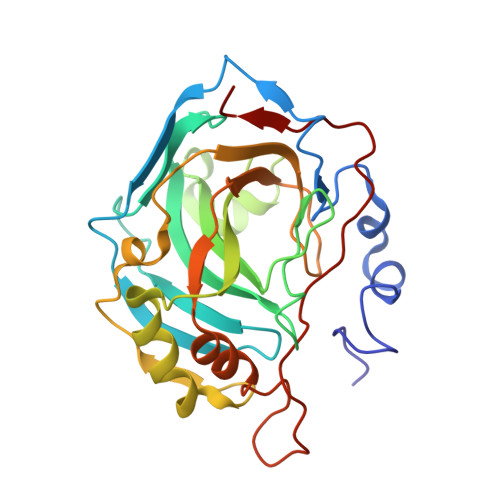
    Indanesulfonamides are interesting lead compounds for designing selective inhibitors of the different isoforms of the zinc enzyme Carbonic Anhydrase (CA). Herein, we report for the first time the X-ray crystal structure of two such derivatives, namely indane-5-sulfonamide and indane-2-valproylamido-5-sulfonamide, in complex with the physiologically dominant human isoform II. The structural analysis reveals that, although these two inhibitors have quite similar chemical structures, the arrangement of their indane ring within the enzyme active site is significantly diverse. Thus, our findings suggest that the introduction of bulky substituents on the indane-sulfonamide ring may alter the binding mode of this potent class of CA inhibitors, although retaining good inhibitory properties. Accordingly, the introduction of bulky tail moieties on the indane-sulfonamide scaffold may represent a powerful strategy to induce a desired physicochemical property to an aromatic sulfonamide or to obtain inhibitors with diverse inhibition profiles and selectivity for various mammalian CAs.


  • Organizational Affiliation
    • Istituto di Biostrutture e Bioimmagini-CNR via Mezzocannone 16, 80134 Napoli, Italy.

Macromolecule Content 

  • Total Structure Weight: 30.07 kDa 
  • Atom Count: 2,508 
  • Modeled Residue Count: 259 
  • Deposited Residue Count: 259 
  • Unique protein chains: 1

Macromolecules

Find similar proteins by:|  3D Structure
Entity ID: 1
MoleculeChains  Sequence LengthOrganismDetailsImage
Carbonic anhydrase 2259Homo sapiensMutation(s): 0 
EC: 4.2.1.1 (PDB Primary Data), 4.2.1.69 (UniProt)
UniProt & NIH Common Fund Data Resources
Find proteins for P00918 (Homo sapiens)
Explore P00918 
Go to UniProtKB:  P00918
PHAROS:  P00918
GTEx:  ENSG00000104267 
Entity Groups
Sequence Clusters30% Identity50% Identity70% Identity90% Identity95% Identity100% Identity
UniProt GroupP00918
Sequence Annotations
Expand
Reference Sequence

Small Molecules

Ligands 4 Unique
IDChains Name / Formula / InChI Key2D Diagram3D Interactions
3CC

Query on 3CC


Download:Ideal Coordinates CCD File
D [auth A]N-[(2R)-5-(aminosulfonyl)-2,3-dihydro-1H-inden-2-yl]-2-propylpentanamide
C17 H26 N2 O3 S
XBYJCVDSFWJBSM-OAHLLOKOSA-N
MBO

Query on MBO


Download:Ideal Coordinates CCD File
C [auth A]MERCURIBENZOIC ACID
C7 H5 Hg O2
FVFZSVRSDNUCGG-UHFFFAOYSA-N
GOL

Query on GOL


Download:Ideal Coordinates CCD File
E [auth A],
F [auth A]
GLYCEROL
C3 H8 O3
PEDCQBHIVMGVHV-UHFFFAOYSA-N
ZN

Query on ZN


Download:Ideal Coordinates CCD File
B [auth A]ZINC ION
Zn
PTFCDOFLOPIGGS-UHFFFAOYSA-N

Experimental Data & Validation

Experimental Data

  • Method: X-RAY DIFFRACTION
  • Resolution: 1.40 Å
  • R-Value Free:  0.199 (Depositor), 0.194 (DCC) 
  • R-Value Work:  0.185 (Depositor), 0.183 (DCC) 
  • R-Value Observed: 0.185 (Depositor) 
Space Group: P 1 21 1
Unit Cell:
Length ( Å )Angle ( ˚ )
a = 42.154α = 90
b = 41.426β = 104.22
c = 71.765γ = 90
Software Package:
Software NamePurpose
CNSrefinement
DENZOdata reduction
SCALEPACKdata scaling
CNSphasing

Structure Validation

View Full Validation Report


Ligand Structure Quality Assessment 


Entry History 

Deposition Data

Revision History  (Full details and data files)

  • Version 1.0: 2008-01-22
    Type: Initial release
  • Version 1.1: 2011-07-13
    Changes: Non-polymer description, Version format compliance
  • Version 1.2: 2023-08-30
    Changes: Data collection, Database references, Derived calculations, Refinement description