Human intestinal maltase-glucoamylase: crystal structure of the N-terminal catalytic subunit and basis of inhibition and substrate specificity
Sim, L., Quezada-Calvillo, R., Sterchi, E.E., Nichols, B.L., Rose, D.R.(2008) J Mol Biology 375: 782-792
- PubMed: 18036614 Search on PubMed
- DOI: https://doi.org/10.1016/j.jmb.2007.10.069
- Primary Citation Related Structures:
2QLY, 2QMJ - PubMed Abstract:
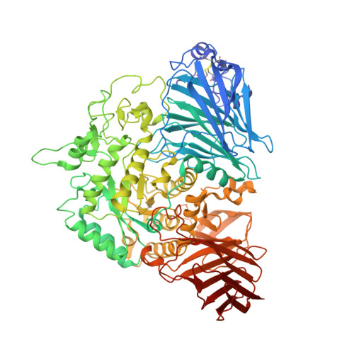
Human maltase-glucoamylase (MGAM) is one of the two enzymes responsible for catalyzing the last glucose-releasing step in starch digestion. MGAM is anchored to the small-intestinal brush-border epithelial cells and contains two homologous glycosyl hydrolase family 31 catalytic subunits: an N-terminal subunit (NtMGAM) found near the membrane-bound end and a C-terminal luminal subunit (CtMGAM). In this study, we report the crystal structure of the human NtMGAM subunit in its apo form (to 2.0 A) and in complex with acarbose (to 1.9 A). Structural analysis of the NtMGAM-acarbose complex reveals that acarbose is bound to the NtMGAM active site primarily through side-chain interactions with its acarvosine unit, and almost no interactions are made with its glycone rings. These observations, along with results from kinetic studies, suggest that the NtMGAM active site contains two primary sugar subsites and that NtMGAM and CtMGAM differ in their substrate specificities despite their structural relationship. Additional sequence analysis of the CtMGAM subunit suggests several features that could explain the higher affinity of the CtMGAM subunit for longer maltose oligosaccharides. The results provide a structural basis for the complementary roles of these glycosyl hydrolase family 31 subunits in the bioprocessing of complex starch structures into glucose.
- Division of Cancer Genomics and Proteomics, Ontario Cancer Institute and Department of Medical Biophysics, University of Toronto, 101 College Street, Toronto, ON, Canada.
Organizational Affiliation: