2QKK | pdb_00002qkk

Human RNase H catalytic domain mutant D210N in complex with 14-mer RNA/DNA hybrid


Experimental Data Snapshot

  • Method: X-RAY DIFFRACTION
  • Resolution: 3.20 Å
  • R-Value Free: 
    0.269 (Depositor), 0.258 (DCC) 
  • R-Value Work: 
    0.212 (Depositor), 0.212 (DCC) 

Starting Model: experimental
View more details

wwPDB Validation 3D Report Full Report

Validation slider image for 2QKK

This is version 1.3 of the entry. See complete history

Literature

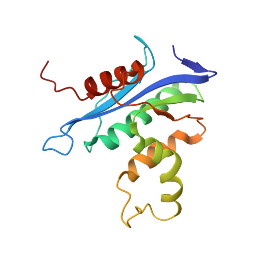
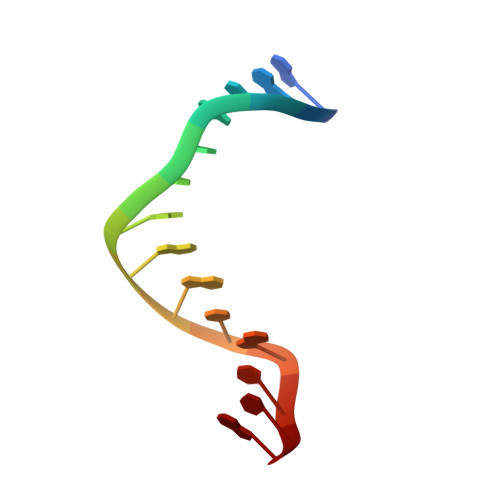
Structure of Human RNase H1 Complexed with an RNA/DNA Hybrid: Insight into HIV Reverse Transcription

Nowotny, M.Gaidamakov, S.A.Ghirlando, R.Cerritelli, S.M.Crouch, R.J.Yang, W.

(2007) Mol Cell 28: 264-276

  • DOI: https://doi.org/10.1016/j.molcel.2007.08.015
  • Primary Citation Related Structures: 
    2QK9, 2QKB, 2QKK

  • PubMed Abstract: 

    We report here crystal structures of human RNase H1 complexed with an RNA/DNA substrate. Unlike B. halodurans RNase H1, human RNase H1 has a basic protrusion, which forms a DNA-binding channel and together with the conserved phosphate-binding pocket confers specificity for the B form and 2'-deoxy DNA. The RNA strand is recognized by four consecutive 2'-OH groups and cleaved by a two-metal ion mechanism. Although RNase H1 is overall positively charged, the substrate interface is neutral to acidic in character, which likely contributes to the catalytic specificity. Positions of the scissile phosphate and two catalytic metal ions are interdependent and highly coupled. Modeling of HIV reverse transcriptase (RT) with RNA/DNA in its RNase H active site suggests that the substrate cannot simultaneously occupy the polymerase active site and must undergo a conformational change to toggle between the two catalytic centers. The region that accommodates this conformational change offers a target to develop HIV-specific inhibitors.


  • Organizational Affiliation
    • Laboratory of Molecular Biology, National Institute of Diabetes and Digestive and Kidney Diseases, National Institutes of Health, Bethesda, MD 20892, USA.

Macromolecule Content 

  • Total Structure Weight: 241.52 kDa 
  • Atom Count: 16,231 
  • Modeled Residue Count: 1,805 
  • Deposited Residue Count: 1,862 
  • Unique protein chains: 1
  • Unique nucleic acid chains: 2

Macromolecules


Find similar proteins by:|  3D Structure
Entity ID: 3
MoleculeChains  Sequence LengthOrganismDetailsImage
Ribonuclease H1154Homo sapiensMutation(s): 1 
Gene Names: RNASEH1RNH1
EC: 3.1.26.4
UniProt & NIH Common Fund Data Resources
Find proteins for O60930 (Homo sapiens)
Explore O60930 
Go to UniProtKB:  O60930
PHAROS:  O60930
GTEx:  ENSG00000171865 
Entity Groups
Sequence Clusters30% Identity50% Identity70% Identity90% Identity95% Identity100% Identity
UniProt GroupO60930
Sequence Annotations
Expand
Reference Sequence
Find similar nucleic acids by:  Sequence
Entity ID: 1
MoleculeChains LengthOrganismImage
5'-R(*CP*GP*AP*CP*AP*CP*CP*UP*GP*AP*UP*UP*CP*C)-3'14N/A
Sequence Annotations
Expand
Reference Sequence
Find similar nucleic acids by:  Sequence
Entity ID: 2
MoleculeChains LengthOrganismImage
5'-D(*GP*GP*AP*AP*TP*CP*AP*GP*GP*TP*GP*TP*CP*G)-3'14N/A
Sequence Annotations
Expand
Reference Sequence

Small Molecules

Ligands 4 Unique
IDChains Name / Formula / InChI Key2D Diagram3D Interactions
MES

Query on MES


Download:Ideal Coordinates CCD File
AA [auth A]2-(N-MORPHOLINO)-ETHANESULFONIC ACID
C6 H13 N O4 S
SXGZJKUKBWWHRA-UHFFFAOYSA-N
TRS

Query on TRS


Download:Ideal Coordinates CCD File
X [auth D]2-AMINO-2-HYDROXYMETHYL-PROPANE-1,3-DIOL
C4 H12 N O3
LENZDBCJOHFCAS-UHFFFAOYSA-O
CA

Query on CA


Download:Ideal Coordinates CCD File
BA [auth B]
DA [auth E]
EA [auth E]
FA [auth F]
GA [auth I]
BA [auth B],
DA [auth E],
EA [auth E],
FA [auth F],
GA [auth I],
HA [auth J],
IA [auth M],
JA [auth M],
KA [auth N],
LA [auth R],
MA [auth S],
NA [auth W],
Y [auth A],
Z [auth A]
CALCIUM ION
Ca
BHPQYMZQTOCNFJ-UHFFFAOYSA-N
CL

Query on CL


Download:Ideal Coordinates CCD File
CA [auth B]CHLORIDE ION
Cl
VEXZGXHMUGYJMC-UHFFFAOYSA-M

Experimental Data & Validation

Experimental Data

  • Method: X-RAY DIFFRACTION
  • Resolution: 3.20 Å
  • R-Value Free:  0.269 (Depositor), 0.258 (DCC) 
  • R-Value Work:  0.212 (Depositor), 0.212 (DCC) 
Space Group: C 1 2 1
Unit Cell:
Length ( Å )Angle ( ˚ )
a = 151.065α = 90
b = 176.2β = 90.22
c = 125.845γ = 90
Software Package:
Software NamePurpose
PHASERphasing
CNSrefinement
HKL-2000data reduction
HKL-2000data scaling

Structure Validation

View Full Validation Report


Entry History 

Deposition Data

Revision History  (Full details and data files)

  • Version 1.0: 2007-11-13
    Type: Initial release
  • Version 1.1: 2011-07-13
    Changes: Version format compliance
  • Version 1.2: 2021-10-20
    Changes: Database references, Derived calculations
  • Version 1.3: 2023-08-30
    Changes: Data collection, Refinement description