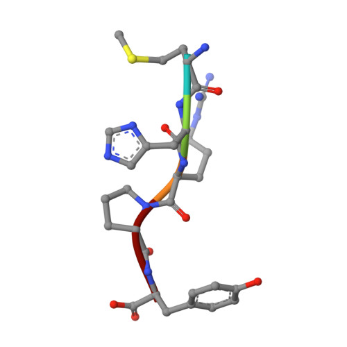
2Q9I

Crystal Structure of D-Dimer from Human Fibrin Complexed with Met-His-Arg-Pro-Tyr-amide.


Experimental Data Snapshot

  • Method: X-RAY DIFFRACTION
  • Resolution: 2.80 Å
  • R-Value Free: 0.276 
  • R-Value Work: 0.216 
  • R-Value Observed: 0.216 

Starting Model: experimental
View more details

wwPDB Validation   3D Report Full Report


This is version 2.1 of the entry. See complete history


Literature

Probing the beta-chain hole of fibrinogen with synthetic peptides that differ at their amino termini

Doolittle, R.F.Pandi, L.

(2007) Biochemistry 46: 10033-10038

  • DOI: https://doi.org/10.1021/bi7010916
  • Primary Citation of Related Structures:  
    2Q9I, 2Z4E

  • PubMed Abstract: 

    In a recent report, we showed that alanine can replace glycine at the amino terminus of synthetic B-knobs that bind to human fibrin(ogen). We now report a survey of 13 synthetic peptides with the general sequence XHRPYam, all tested with regard to their ability to delay fibrinolysis in an in vitro system activated by t-PA, the results being used as measures of binding affinity to the betaC hole. Unexpectedly, some large and bulky amino acids, including methionine and arginine, are effective binders. Amino acids that branch at the beta carbon (valine, isoleucine, and threonine) do not bind effectively. Crystal structures were determined for two of the peptides (GHRPYam and MHRPYam) complexed with fibrin fragment D-dimer; the modeling of various other side chains showed clashing in the cases of beta-carbon substituents. The two crystal structures also showed that the enhanced binding observed with pentapeptides with carboxyl-terminal tyrosine, compared with that of their tetrapeptide equivalents, is attributable to an interaction between the tyrosine side chain and a guanidino group of a nearby arginine (beta406). The equivalent position in gamma-chains of human fibrin(ogen) is occupied by a lysine (gamma338), but in chicken and lamprey fibrin(ogen), it is an arginine, just as occurs in beta chains. Accordingly, the peptides GPRPam and GPRPYam, which are surrogate A-knobs, were tested for their influence on fibrin polymerization with fibrinogen from lamprey and humans. In lampreys, GPRPYam is a significantly better inhibitor, but in humans, it is less effective than GPRPam, indicating that in the lamprey system the same tyrosine-arginine interaction can also occur in the gamma-chain setting.


  • Organizational Affiliation

    Department of Chemistry and Biochemistry and Division of Biology, University of California, San Diego, La Jolla, California 92093-0314, USA. rdoolittle@ucsd.edu


Macromolecules
Find similar proteins by:  (by identity cutoff)  |  3D Structure
Entity ID: 1
MoleculeChains Sequence LengthOrganismDetailsImage
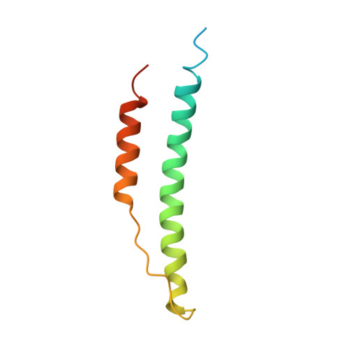
Fibrinogen alpha chain
A, D
87Homo sapiensMutation(s): 0 
UniProt & NIH Common Fund Data Resources
Find proteins for P02671 (Homo sapiens)
Explore P02671 
Go to UniProtKB:  P02671
PHAROS:  P02671
GTEx:  ENSG00000171560 
Entity Groups  
Sequence Clusters30% Identity50% Identity70% Identity90% Identity95% Identity100% Identity
UniProt GroupP02671
Sequence Annotations
Expand
  • Reference Sequence
Find similar proteins by:  (by identity cutoff)  |  3D Structure
Entity ID: 2
MoleculeChains Sequence LengthOrganismDetailsImage
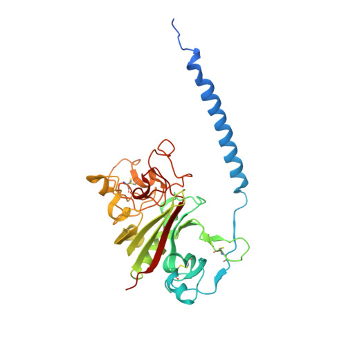
Fibrinogen beta chain
B, E
328Homo sapiensMutation(s): 0 
UniProt & NIH Common Fund Data Resources
Find proteins for P02675 (Homo sapiens)
Explore P02675 
Go to UniProtKB:  P02675
PHAROS:  P02675
GTEx:  ENSG00000171564 
Entity Groups  
Sequence Clusters30% Identity50% Identity70% Identity90% Identity95% Identity100% Identity
UniProt GroupP02675
Glycosylation
Glycosylation Sites: 1Go to GlyGen: P02675-1
Sequence Annotations
Expand
  • Reference Sequence
Find similar proteins by:  (by identity cutoff)  |  3D Structure
Entity ID: 3
MoleculeChains Sequence LengthOrganismDetailsImage
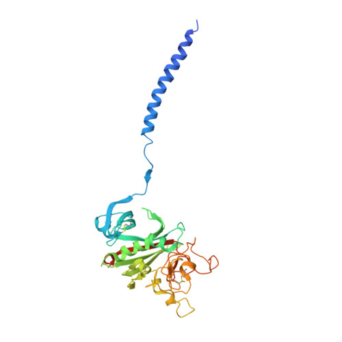
Fibrinogen, gamma polypeptide
C, F
324Homo sapiensMutation(s): 0 
UniProt & NIH Common Fund Data Resources
Find proteins for P02679 (Homo sapiens)
Explore P02679 
Go to UniProtKB:  P02679
PHAROS:  P02679
GTEx:  ENSG00000171557 
Entity Groups  
Sequence Clusters30% Identity50% Identity70% Identity90% Identity95% Identity100% Identity
UniProt GroupP02679
Sequence Annotations
Expand
  • Reference Sequence

Find similar proteins by:  Sequence   |   3D Structure  

Entity ID: 4
MoleculeChains Sequence LengthOrganismDetailsImage
Fibrin B Knob (GHRPam)G [auth S],
H [auth T]
4N/AMutation(s): 0 
Sequence Annotations
Expand
  • Reference Sequence

Find similar proteins by:  Sequence   |   3D Structure  

Entity ID: 5
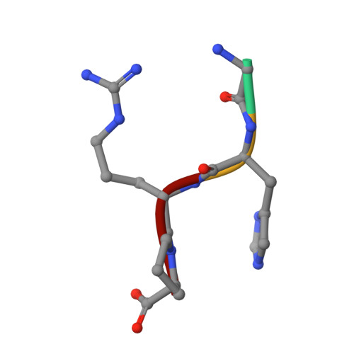
MoleculeChains Sequence LengthOrganismDetailsImage
Fibrin B Knob (MHRPYam)I [auth M],
J [auth N]
5N/AMutation(s): 0 
Sequence Annotations
Expand
  • Reference Sequence
Oligosaccharides

Help

Entity ID: 6
MoleculeChains Length2D Diagram Glycosylation3D Interactions
2-acetamido-2-deoxy-beta-D-glucopyranose-(1-4)-2-acetamido-2-deoxy-beta-D-glucopyranoseK [auth G],
L [auth H]
2N-Glycosylation
Glycosylation Resources
GlyTouCan:  G42666HT
GlyCosmos:  G42666HT
GlyGen:  G42666HT
Experimental Data & Validation

Experimental Data

  • Method: X-RAY DIFFRACTION
  • Resolution: 2.80 Å
  • R-Value Free: 0.276 
  • R-Value Work: 0.216 
  • R-Value Observed: 0.216 
  • Space Group: P 21 21 21
Unit Cell:
Length ( Å )Angle ( ˚ )
a = 54.38α = 90
b = 147.23β = 90
c = 231.48γ = 90
Software Package:
Software NamePurpose
CNSrefinement
PDB_EXTRACTdata extraction
MAR345dtbdata collection
DENZOdata reduction
SCALEPACKdata scaling
CNSphasing

Structure Validation

View Full Validation Report


Entry History 

Deposition Data

Revision History  (Full details and data files)

  • Version 1.0: 2007-12-11
    Type: Initial release
  • Version 1.1: 2011-07-13
    Changes: Non-polymer description, Version format compliance
  • Version 2.0: 2020-07-29
    Type: Remediation
    Reason: Carbohydrate remediation
    Changes: Atomic model, Data collection, Derived calculations, Structure summary
  • Version 2.1: 2023-08-30
    Changes: Data collection, Database references, Refinement description, Structure summary