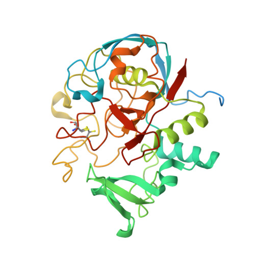
Function and structure of a prokaryotic formylglycine-generating enzyme.
Carlson, B.L., Ballister, E.R., Skordalakes, E., King, D.S., Breidenbach, M.A., Gilmore, S.A., Berger, J.M., Bertozzi, C.R.(2008) J Biol Chem 283: 20117-20125
- PubMed: 18390551 Search on PubMedSearch on PubMed Central
- DOI: https://doi.org/10.1074/jbc.M800217200
- Primary Citation Related Structures:
2Q17 - PubMed Abstract:
Type I sulfatases require an unusual co- or post-translational modification for their activity in hydrolyzing sulfate esters. In eukaryotic sulfatases, an active site cysteine residue is oxidized to the aldehyde-containing C(alpha)-formylglycine residue by the formylglycine-generating enzyme (FGE). The machinery responsible for sulfatase activation is poorly understood in prokaryotes. Here we describe the identification of a prokaryotic FGE from Mycobacterium tuberculosis. In addition, we solved the crystal structure of the Streptomyces coelicolor FGE homolog to 2.1 A resolution. The prokaryotic homolog exhibits remarkable structural similarity to human FGE, including the position of catalytic cysteine residues. Both biochemical and structural data indicate the presence of an oxidized cysteine modification in the active site that may be relevant to catalysis. In addition, we generated a mutant M. tuberculosis strain lacking FGE. Although global sulfatase activity was reduced in the mutant, a significant amount of residual sulfatase activity suggests the presence of FGE-independent sulfatases in this organism.
- Department of Molecular and Cell Biology, Howard Hughes Medical Institute, University of California Berkeley, Berkeley, CA 94720, USA.
Organizational Affiliation: