2Q0S

Structure of the Inhibitor bound form of M. Smegmatis Aryl Esterase


Experimental Data Snapshot

  • Method: X-RAY DIFFRACTION
  • Resolution: 1.50 Å
  • R-Value Free: 0.160 
  • R-Value Work: 0.134 
  • R-Value Observed: 0.135 

wwPDB Validation   3D Report Full Report


This is version 1.2 of the entry. See complete history


Literature

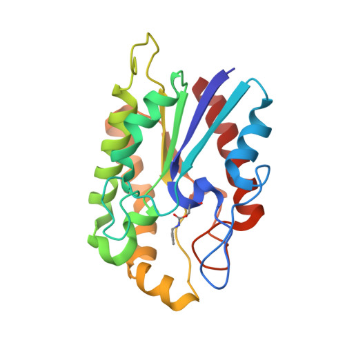
Structure of a novel enzyme that catalyzes acyl transfer to alcohols in aqueous conditions.

Mathews, I.Soltis, M.Saldajeno, M.Ganshaw, G.Sala, R.Weyler, W.Cervin, M.A.Whited, G.Bott, R.

(2007) Biochemistry 46: 8969-8979

  • DOI: https://doi.org/10.1021/bi7002444
  • Primary Citation of Related Structures:  
    2Q0Q, 2Q0S

  • PubMed Abstract: 

    The unusual architecture of the enzyme (MsAcT) isolated from Mycobacterium smegmatis forms the mechanistic basis for favoring alcoholysis over hydrolysis in water. Unlike hydrolases that perform alcoholysis only under anhydrous conditions, MsAcT demonstrates alcoholysis in substantially aqueous media and, in the presence of hydrogen peroxide, has a perhydrolysis:hydrolysis ratio 50-fold greater than that of the best lipase tested. The crystal structures of the apoenzyme and an inhibitor-bound form have been determined to 1.5 A resolution. MsAcT is an octamer in the asymmetric unit and forms a tightly associated aggregate in solution. Relative to other structurally similar monomers, MsAcT contains several insertions that contribute to the oligomerization and greatly restrict the shape of the active site, thereby limiting its accessibility. These properties create an environment by which MsAcT can catalyze transesterification reactions in an aqueous medium and suggests how a serine hydrolase can be engineered to be an efficient acyltransferase.


  • Organizational Affiliation

    Stanford Synchrotron Research Laboratory, 2575 Sand Hill Road, Menlo Park, California 94025, USA.


Macromolecules
Find similar proteins by:  (by identity cutoff)  |  3D Structure
Entity ID: 1
MoleculeChains Sequence LengthOrganismDetailsImage
Aryl esterase
A, B, C, D, E
A, B, C, D, E, F, G, H
216Mycolicibacterium smegmatisMutation(s): 1 
EC: 3.1.1.2
UniProt
Find proteins for A0R5U7 (Mycolicibacterium smegmatis (strain ATCC 700084 / mc(2)155))
Explore A0R5U7 
Go to UniProtKB:  A0R5U7
Entity Groups  
Sequence Clusters30% Identity50% Identity70% Identity90% Identity95% Identity100% Identity
UniProt GroupA0R5U7
Sequence Annotations
Expand
  • Reference Sequence
Small Molecules
Ligands 1 Unique
IDChains Name / Formula / InChI Key2D Diagram3D Interactions
SO4
Query on SO4

Download Ideal Coordinates CCD File 
I [auth B]SULFATE ION
O4 S
QAOWNCQODCNURD-UHFFFAOYSA-L
Modified Residues  1 Unique
IDChains TypeFormula2D DiagramParent
N10
Query on N10
A, B, C, D, E
A, B, C, D, E, F, G, H
L-PEPTIDE LINKINGC10 H20 N2 O4SER
Experimental Data & Validation

Experimental Data

  • Method: X-RAY DIFFRACTION
  • Resolution: 1.50 Å
  • R-Value Free: 0.160 
  • R-Value Work: 0.134 
  • R-Value Observed: 0.135 
  • Space Group: P 1
Unit Cell:
Length ( Å )Angle ( ˚ )
a = 67.754α = 104.1
b = 80.096β = 112.1
c = 85.974γ = 97.4
Software Package:
Software NamePurpose
REFMACrefinement
ADSCdata collection
HKL-2000data reduction
SCALEPACKdata scaling
MOLREPphasing

Structure Validation

View Full Validation Report


Entry History 

Deposition Data

Revision History  (Full details and data files)

  • Version 1.0: 2007-12-11
    Type: Initial release
  • Version 1.1: 2011-07-13
    Changes: Version format compliance
  • Version 1.2: 2024-04-03
    Changes: Data collection, Database references, Derived calculations, Refinement description