


Crystal structure of E. coli phosphoenolpyruvate carboxykinase (PEPCK) complexed with ATP, Mg2+, Mn2+, carbon dioxide and oxaloacetate
Delbaere, L.T.J., Cotelesage, J.J.H., Goldie, H.To be published.
Experimental Data Snapshot
Entity ID: 1 | |||||
|---|---|---|---|---|---|
| Molecule | Chains | Sequence Length | Organism | Details | Image |
| Phosphoenolpyruvate carboxykinase | A [auth X] | 540 | Escherichia coli K-12 | Mutation(s): 0 Gene Names: pckA EC: 4.1.1.49 | 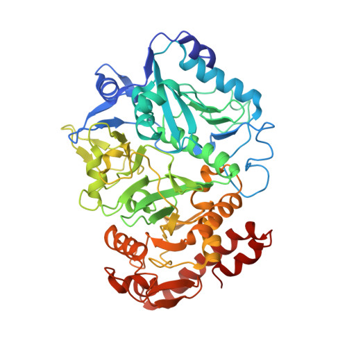 |
UniProt | |||||
Entity Groups | |||||
| Sequence Clusters | 30% Identity50% Identity70% Identity90% Identity95% Identity100% Identity | ||||
| UniProt Group | P22259 | ||||
Sequence AnnotationsExpand | |||||
Reference Sequence | |||||
| Ligands 5 Unique | |||||
|---|---|---|---|---|---|
| ID | Chains | Name / Formula / InChI Key | 2D Diagram | 3D Interactions | |
| ATP Download:Ideal Coordinates CCD File | E [auth X] | ADENOSINE-5'-TRIPHOSPHATE C10 H16 N5 O13 P3 ZKHQWZAMYRWXGA-KQYNXXCUSA-N |  | ||
| OAA Download:Ideal Coordinates CCD File | D [auth X] | OXALOACETATE ION C4 H3 O5 KHPXUQMNIQBQEV-UHFFFAOYSA-M |  | ||
| MN Download:Ideal Coordinates CCD File | C [auth X] | MANGANESE (II) ION Mn WAEMQWOKJMHJLA-UHFFFAOYSA-N |  | ||
| CO2 Download:Ideal Coordinates CCD File | F [auth X] | CARBON DIOXIDE C O2 CURLTUGMZLYLDI-UHFFFAOYSA-N |  | ||
| MG Download:Ideal Coordinates CCD File | B [auth X] | MAGNESIUM ION Mg JLVVSXFLKOJNIY-UHFFFAOYSA-N |  | ||
| Length ( Å ) | Angle ( ˚ ) |
|---|---|
| a = 124.556 | α = 90 |
| b = 94.302 | β = 95.54 |
| c = 46.354 | γ = 90 |
| Software Name | Purpose |
|---|---|
| REFMAC | refinement |
| MOSFLM | data reduction |
| SCALA | data scaling |
| PHASER | phasing |


