2PQY | pdb_00002pqy

E. coli RNase 1 (in vitro refolded with DsbA only)


Experimental Data Snapshot

  • Method: X-RAY DIFFRACTION
  • Resolution: 2.30 Å
  • R-Value Free: 
    0.240 (Depositor), 0.240 (DCC) 
  • R-Value Work: 
    0.196 (Depositor), 0.190 (DCC) 

Starting Model: experimental
View more details

wwPDB Validation   3D Report Full Report


This is version 1.4 of the entry. See complete history


Literature

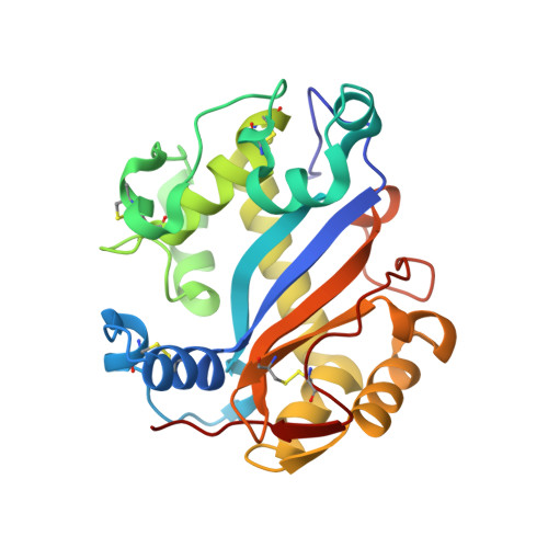
The oxidase DsbA folds a protein with a nonconsecutive disulfide.

Messens, J.Collet, J.F.Van Belle, K.Brosens, E.Loris, R.Wyns, L.

(2007) J Biological Chem 282: 31302-31307

  • DOI: https://doi.org/10.1074/jbc.M705236200
  • Primary Citation of Related Structures:  
    2PQY

  • PubMed Abstract: 

    One of the last unsolved problems of molecular biology is how the sequential amino acid information leads to a functional protein. Correct disulfide formation within a protein is hereby essential. We present periplasmic ribonuclease I (RNase I) from Escherichia coli as a new endogenous substrate for the study of oxidative protein folding. One of its four disulfides is between nonconsecutive cysteines. In general view, the folding of proteins with nonconsecutive disulfides requires the protein disulfide isomerase DsbC. In contrast, our study with RNase I shows that DsbA is a sufficient catalyst for correct disulfide formation in vivo and in vitro. DsbA is therefore more specific than generally assumed. Further, we show that the redox potential of the periplasm depends on the presence of glutathione and the Dsb proteins to maintain it at-165 mV. We determined the influence of this redox potential on the folding of RNase I. Under the more oxidizing conditions of dsb(-) strains, DsbC becomes necessary to correct non-native disulfides, but it cannot substitute for DsbA. Altogether, DsbA folds a protein with a nonconsecutive disulfide as long as no incorrect disulfides are formed.


  • Organizational Affiliation
    • Brussels Center for Redox Biology, Vlaams Instituut voor Biotechnologie, Vrije Universiteit Brussel, 1050 Brussel, Belgium. joris.messens@vub.ac.be

Macromolecules
Find similar proteins by:  (by identity cutoff)  |  3D Structure
Entity ID: 1
MoleculeChains Sequence LengthOrganismDetailsImage
Ribonuclease I245Escherichia coliMutation(s): 0 
Gene Names: rnarnsA
EC: 3.1.27.6 (PDB Primary Data), 4.6.1.21 (UniProt)
UniProt
Find proteins for P21338 (Escherichia coli (strain K12))
Explore P21338 
Go to UniProtKB:  P21338
Entity Groups  
Sequence Clusters30% Identity50% Identity70% Identity90% Identity95% Identity100% Identity
UniProt GroupP21338
Sequence Annotations
Expand
  • Reference Sequence
Experimental Data & Validation

Experimental Data

  • Method: X-RAY DIFFRACTION
  • Resolution: 2.30 Å
  • R-Value Free:  0.240 (Depositor), 0.240 (DCC) 
  • R-Value Work:  0.196 (Depositor), 0.190 (DCC) 
Space Group: P 1 21 1
Unit Cell:
Length ( Å )Angle ( ˚ )
a = 39.571α = 90
b = 49.548β = 97.682
c = 53.328γ = 90
Software Package:
Software NamePurpose
MAR345data collection
CNSrefinement
DENZOdata reduction
SCALEPACKdata scaling

Structure Validation

View Full Validation Report


Entry History 

Deposition Data

Revision History  (Full details and data files)

  • Version 1.0: 2007-08-14
    Type: Initial release
  • Version 1.1: 2011-07-13
    Changes: Version format compliance
  • Version 1.2: 2017-10-18
    Changes: Refinement description
  • Version 1.3: 2023-08-30
    Changes: Data collection, Database references, Derived calculations, Refinement description
  • Version 1.4: 2024-10-30
    Changes: Structure summary