2PQA

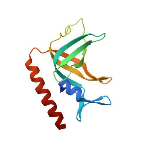
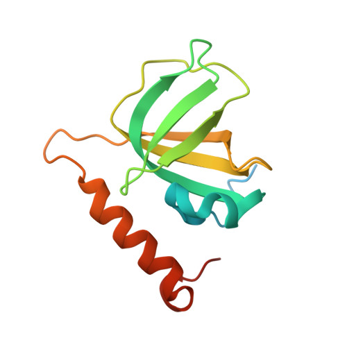
Crystal Structure of Full-length Human RPA 14/32 Heterodimer


Experimental Data Snapshot

  • Method: X-RAY DIFFRACTION
  • Resolution: 2.50 Å
  • R-Value Free: 0.280 
  • R-Value Work: 0.228 
  • R-Value Observed: 0.231 

wwPDB Validation   3D Report Full Report


This is version 1.2 of the entry. See complete history


Literature

Structure of the full-length human RPA14/32 complex gives insights into the mechanism of DNA binding and complex formation.

Deng, X.Habel, J.E.Kabaleeswaran, V.Snell, E.H.Wold, M.S.Borgstahl, G.E.

(2007) J Mol Biol 374: 865-876

  • DOI: https://doi.org/10.1016/j.jmb.2007.09.074
  • Primary Citation of Related Structures:  
    2PI2, 2PQA, 2Z6K

  • PubMed Abstract: 

    Replication protein A (RPA) is the ubiquitous, eukaryotic single-stranded DNA (ssDNA) binding protein and is essential for DNA replication, recombination, and repair. Here, crystal structures of the soluble RPA heterodimer, composed of the RPA14 and RPA32 subunits, have been determined for the full-length protein in multiple crystal forms. In all crystals, the electron density for the N-terminal (residues 1-42) and C-terminal (residues 175-270) regions of RPA32 is weak and of poor quality indicating that these regions are disordered and/or assume multiple positions in the crystals. Hence, the RPA32 N terminus, that is hyperphosphorylated in a cell-cycle-dependent manner and in response to DNA damaging agents, appears to be inherently disordered in the unphosphorylated state. The C-terminal, winged helix-loop-helix, protein-protein interaction domain adopts several conformations perhaps to facilitate its interaction with various proteins. Although the ordered regions of RPA14/32 resemble the previously solved protease-resistant core crystal structure, the quaternary structures between the heterodimers are quite different. Thus, the four-helix bundle quaternary assembly noted in the original core structure is unlikely to be related to the quaternary structure of the intact heterotrimer. An organic ligand binding site between subunits RPA14 and RPA32 was identified to bind dioxane. Comparison of the ssDNA binding surfaces of RPA70 with RPA14/32 showed that the lower affinity of RPA14/32 can be attributed to a shallower binding crevice with reduced positive electrostatic charge.


  • Organizational Affiliation

    The Eppley Institute for Research in Cancer and Allied Diseases, 987696 Nebraska Medical Center, Omaha, NE 68198-7696, USA.


Macromolecules
Find similar proteins by:  (by identity cutoff)  |  3D Structure
Entity ID: 1
MoleculeChains Sequence LengthOrganismDetailsImage
Replication protein A 32 kDa subunit
A, C
131Homo sapiensMutation(s): 0 
Gene Names: RPA2REPA2RPA32
UniProt & NIH Common Fund Data Resources
Find proteins for P15927 (Homo sapiens)
Explore P15927 
Go to UniProtKB:  P15927
PHAROS:  P15927
GTEx:  ENSG00000117748 
Entity Groups  
Sequence Clusters30% Identity50% Identity70% Identity90% Identity95% Identity100% Identity
UniProt GroupP15927
Sequence Annotations
Expand
  • Reference Sequence
Find similar proteins by:  (by identity cutoff)  |  3D Structure
Entity ID: 2
MoleculeChains Sequence LengthOrganismDetailsImage
Replication protein A 14 kDa subunit
B, D
142Homo sapiensMutation(s): 0 
Gene Names: RPA3REPA3RPA14
UniProt & NIH Common Fund Data Resources
Find proteins for P35244 (Homo sapiens)
Explore P35244 
Go to UniProtKB:  P35244
PHAROS:  P35244
GTEx:  ENSG00000106399 
Entity Groups  
Sequence Clusters30% Identity50% Identity70% Identity90% Identity95% Identity100% Identity
UniProt GroupP35244
Sequence Annotations
Expand
  • Reference Sequence
Experimental Data & Validation

Experimental Data

  • Method: X-RAY DIFFRACTION
  • Resolution: 2.50 Å
  • R-Value Free: 0.280 
  • R-Value Work: 0.228 
  • R-Value Observed: 0.231 
  • Space Group: P 65
Unit Cell:
Length ( Å )Angle ( ˚ )
a = 63.377α = 90
b = 63.377β = 90
c = 272.641γ = 120
Software Package:
Software NamePurpose
REFMACrefinement
HKL-2000data collection
HKL-2000data reduction
HKL-2000data scaling
SHELXSphasing

Structure Validation

View Full Validation Report


Entry History 

Deposition Data

Revision History  (Full details and data files)

  • Version 1.0: 2007-11-13
    Type: Initial release
  • Version 1.1: 2011-07-13
    Changes: Derived calculations, Version format compliance
  • Version 1.2: 2024-02-21
    Changes: Data collection, Database references