2PHK

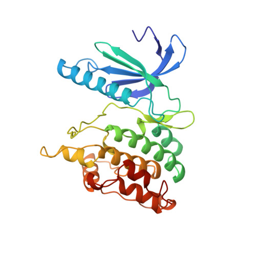
THE CRYSTAL STRUCTURE OF A PHOSPHORYLASE KINASE PEPTIDE SUBSTRATE COMPLEX: KINASE SUBSTRATE RECOGNITION


Experimental Data Snapshot

  • Method: X-RAY DIFFRACTION
  • Resolution: 2.60 Å
  • R-Value Free: 0.300 
  • R-Value Work: 0.236 
  • R-Value Observed: 0.253 

Starting Model: experimental
View more details

wwPDB Validation   3D Report Full Report


This is version 1.6 of the entry. See complete history


Literature

The crystal structure of a phosphorylase kinase peptide substrate complex: kinase substrate recognition.

Lowe, E.D.Noble, M.E.Skamnaki, V.T.Oikonomakos, N.G.Owen, D.J.Johnson, L.N.

(1997) EMBO J 16: 6646-6658

  • DOI: https://doi.org/10.1093/emboj/16.22.6646
  • Primary Citation of Related Structures:  
    2PHK

  • PubMed Abstract: 

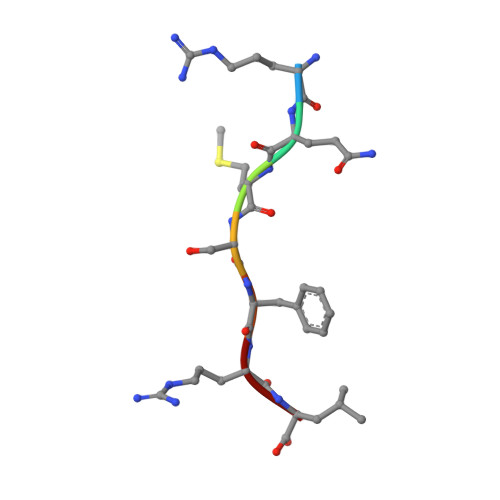
    The structure of a truncated form of the gamma-subunit of phosphorylase kinase (PHKgammat) has been solved in a ternary complex with a non-hydrolysable ATP analogue (adenylyl imidodiphosphate, AMPPNP) and a heptapeptide substrate related in sequence to both the natural substrate and to the optimal peptide substrate. Kinetic characterization of the phosphotransfer reaction confirms the peptide to be a good substrate, and the structure allows identification of key features responsible for its high affinity. Unexpectedly, the substrate peptide forms a short anti-parallel beta-sheet with the kinase activation segment, the region which in other kinases plays an important role in regulation of enzyme activity. This anchoring of the main chain of the substrate peptide at a fixed distance from the gamma-phosphate of ATP explains the selectivity of PHK for serine/threonine over tyrosine as a substrate. The catalytic core of PHK exists as a dimer in crystals of the ternary complex, and the relevance of this phenomenon to its in vivo recognition of dimeric glycogen phosphorylase b is considered.


  • Organizational Affiliation

    Laboratory of Molecular Biophysics and Oxford Centre for Molecular Sciences, University of Oxford, Rex Richards Building, South Parks Road, Oxford OX1 3QU, UK.


Macromolecules
Find similar proteins by:  (by identity cutoff)  |  3D Structure
Entity ID: 1
MoleculeChains Sequence LengthOrganismDetailsImage
PHOSPHORYLASE KINASE277Oryctolagus cuniculusMutation(s): 0 
Gene Names: PHKG
EC: 2.7.1.38
UniProt
Find proteins for P00518 (Oryctolagus cuniculus)
Explore P00518 
Go to UniProtKB:  P00518
Entity Groups  
Sequence Clusters30% Identity50% Identity70% Identity90% Identity95% Identity100% Identity
UniProt GroupP00518
Sequence Annotations
Expand
  • Reference Sequence

Find similar proteins by:  Sequence   |   3D Structure  

Entity ID: 2
MoleculeChains Sequence LengthOrganismDetailsImage
MC-PEPTIDE7N/AMutation(s): 0 
Sequence Annotations
Expand
  • Reference Sequence
Small Molecules
Ligands 3 Unique
IDChains Name / Formula / InChI Key2D Diagram3D Interactions
ATP
Query on ATP

Download Ideal Coordinates CCD File 
E [auth A]ADENOSINE-5'-TRIPHOSPHATE
C10 H16 N5 O13 P3
ZKHQWZAMYRWXGA-KQYNXXCUSA-N
GOL
Query on GOL

Download Ideal Coordinates CCD File 
F [auth A]GLYCEROL
C3 H8 O3
PEDCQBHIVMGVHV-UHFFFAOYSA-N
MN
Query on MN

Download Ideal Coordinates CCD File 
C [auth A],
D [auth A]
MANGANESE (II) ION
Mn
WAEMQWOKJMHJLA-UHFFFAOYSA-N
Experimental Data & Validation

Experimental Data

  • Method: X-RAY DIFFRACTION
  • Resolution: 2.60 Å
  • R-Value Free: 0.300 
  • R-Value Work: 0.236 
  • R-Value Observed: 0.253 
  • Space Group: P 32 2 1
Unit Cell:
Length ( Å )Angle ( ˚ )
a = 65.3α = 90
b = 65.3β = 90
c = 145.8γ = 120
Software Package:
Software NamePurpose
AMoREphasing
REFMACrefinement
MOSFLMdata reduction
CCP4data scaling

Structure Validation

View Full Validation Report


Entry History 

Deposition Data

Revision History  (Full details and data files)

  • Version 1.0: 1999-01-13
    Type: Initial release
  • Version 1.1: 2008-03-03
    Changes: Version format compliance
  • Version 1.2: 2011-07-13
    Changes: Non-polymer description, Version format compliance
  • Version 1.3: 2018-04-04
    Changes: Data collection
  • Version 1.4: 2018-04-11
    Changes: Data collection
  • Version 1.5: 2023-08-09
    Changes: Database references, Derived calculations, Refinement description
  • Version 1.6: 2024-05-22
    Changes: Data collection