2PG8

Crystal structure of R254K mutanat of DpgC with bound substrate analog


Experimental Data Snapshot

  • Method: X-RAY DIFFRACTION
  • Resolution: 3.00 Å
  • R-Value Free: 0.367 
  • R-Value Work: 0.331 

wwPDB Validation   3D Report Full Report


Ligand Structure Quality Assessment 


This is version 1.4 of the entry. See complete history


Literature

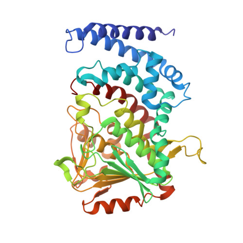
Substrate Recognition and Catalysis by the Cofactor-Independent Dioxygenase DpgC.

Fielding, E.N.Widboom, P.F.Bruner, S.D.

(2007) Biochemistry 46: 13994-14000

  • DOI: https://doi.org/10.1021/bi701148b
  • Primary Citation of Related Structures:  
    2PG8

  • PubMed Abstract: 

    The enzyme DpgC belongs to a small class of oxygenases not dependent on accessory cofactors for activity. DpgC is in the biosynthetic pathway for the nonproteinogenic amino acid 3,5-dihydroxyphenylglycine in actinomycetes bacteria responsible for the production of the vancomycin/teicoplanin family of antibiotic natural products. The X-ray structure of DpgC [Widboom, P. W., Fielding, E. N., Liu, Y., and Bruner, S. D. (2007) Nature 447, 342-345] confirmed the absence of cofactors and defined a novel hydrophobic dioxygen binding pocket adjacent to a bound substrate analogue. In this paper, the role specific amino acids play in substrate recognition and catalysis is examined through biochemical and structural characterization of site-specific enzyme mutations and alternate substrates. The results establish the importance of three amino acids, Arg254, Glu299, and Glu189, in the chemistry of DpgC. Arg254 and Glu189 join to form a specific contact with one of the phenolic hydroxyls of the substrate, and this interaction plays a key role in both substrate recognition and catalysis. The X-ray crystal structure of Arg254Lys was determined to address the role this residue plays in the chemistry. In addition, characterization of alternate substrate analogues demonstrates the presence and position of phenol groups are necessary for both enzyme recognition and downstream oxidation chemistry. Overall, this work defines the mechanism of substrate recognition and specificity by the cofactor-independent dioxygenase DpgC.


  • Organizational Affiliation

    Department of Chemistry, Merkert Chemistry Center, Boston College, Chestnut Hill, Massachusetts 02467, USA.


Macromolecules
Find similar proteins by:  (by identity cutoff)  |  3D Structure
Entity ID: 1
MoleculeChains Sequence LengthOrganismDetailsImage
DpgC
A, B, C
417Streptomyces toyocaensisMutation(s): 1 
Gene Names: DpgC
UniProt
Find proteins for Q8KLK7 (Streptomyces toyocaensis)
Explore Q8KLK7 
Go to UniProtKB:  Q8KLK7
Entity Groups  
Sequence Clusters30% Identity50% Identity70% Identity90% Identity95% Identity100% Identity
UniProt GroupQ8KLK7
Sequence Annotations
Expand
  • Reference Sequence
Small Molecules
Ligands 2 Unique
IDChains Name / Formula / InChI Key2D Diagram3D Interactions
YE1
Query on YE1

Download Ideal Coordinates CCD File 
D [auth A],
F [auth B],
G [auth C]
[(2R,3S,4R,5R)-5-(6-AMINO-9H-PURIN-9-YL)-4-HYDROXY-3-(PHOSPHONOOXY)TETRAHYDROFURAN-2-YL]METHYL (3R)-4-({3-[(2-{[(3,5-DIHYDROXYPHENYL)ACETYL]AMINO}ETHYL)AMINO]-3-OXOPROPYL}AMINO)-3-HYDROXY-2,2-DIMETHYL-4-OXOBUTYL DIHYDROGEN DIPHOSPHATE
C29 H43 N8 O19 P3
MSJGSRHUBFYIJV-CECATXLMSA-N
OXY
Query on OXY

Download Ideal Coordinates CCD File 
E [auth A],
H [auth C]
OXYGEN MOLECULE
O2
MYMOFIZGZYHOMD-UHFFFAOYSA-N
Experimental Data & Validation

Experimental Data

  • Method: X-RAY DIFFRACTION
  • Resolution: 3.00 Å
  • R-Value Free: 0.367 
  • R-Value Work: 0.331 
  • Space Group: P 21 21 2
Unit Cell:
Length ( Å )Angle ( ˚ )
a = 139.054α = 90
b = 155.309β = 90
c = 169.208γ = 90
Software Package:
Software NamePurpose
DENZOdata reduction
SCALEPACKdata scaling
CNSrefinement
REFMACrefinement
PDB_EXTRACTdata extraction
HKL-2000data collection
HKL-2000data reduction
CNSphasing

Structure Validation

View Full Validation Report


Ligand Structure Quality Assessment 


Entry History 

Deposition Data

Revision History  (Full details and data files)

  • Version 1.0: 2008-01-22
    Type: Initial release
  • Version 1.1: 2011-07-13
    Changes: Derived calculations, Version format compliance
  • Version 1.2: 2017-10-18
    Changes: Refinement description
  • Version 1.3: 2021-10-20
    Changes: Database references, Derived calculations
  • Version 1.4: 2023-08-30
    Changes: Data collection, Refinement description