2PFJ | pdb_00002pfj

Crystal Structure of T7 Endo I resolvase in complex with a Holliday Junction


Experimental Data Snapshot

  • Method: X-RAY DIFFRACTION
  • Resolution: 3.10 Å
  • R-Value Free: 
    0.298 (Depositor), 0.291 (DCC) 
  • R-Value Work: 
    0.267 (Depositor), 0.262 (DCC) 
  • R-Value Observed: 
    0.268 (Depositor) 

Starting Model: experimental
View more details

wwPDB Validation 3D Report Full Report

Validation slider image for 2PFJ

This is version 1.3 of the entry. See complete history

Literature

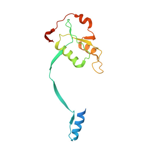
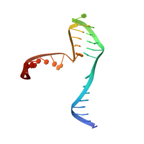
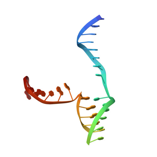
The structural basis of Holliday junction resolution by T7 endonuclease I.

Hadden, J.M.Declais, A.C.Carr, S.B.Lilley, D.M.Phillips, S.E.

(2007) Nature 449: 621-624

  • DOI: https://doi.org/10.1038/nature06158
  • Primary Citation Related Structures: 
    2PFJ

  • PubMed Abstract: 

    The four-way (Holliday) DNA junction is the central intermediate in homologous recombination, a ubiquitous process that is important in DNA repair and generation of genetic diversity. The penultimate stage of recombination requires resolution of the DNA junction into nicked-duplex species by the action of a junction-resolving enzyme, examples of which have been identified in a wide variety of organisms. These enzymes are nucleases that are highly selective for the structure of branched DNA. The mechanism of this selectivity has, however, been unclear in the absence of structural data. Here we present the crystal structure of the junction-resolving enzyme phage T7 endonuclease I in complex with a synthetic four-way DNA junction. Although the enzyme is structure-selective, significant induced fit occurs in the interaction, with changes in the structure of both the protein and the junction. The dimeric enzyme presents two binding channels that contact the backbones of the junction's helical arms over seven nucleotides. These interactions effectively measure the relative orientations and positions of the arms of the junction, thereby ensuring that binding is selective for branched DNA that can achieve this geometry.


  • Organizational Affiliation
    • Astbury Centre for Structural Molecular Biology, Institute of Molecular and Cellular Biology, Faculty of Biological Sciences, University of Leeds, Leeds LS2 9JT, UK.

Macromolecule Content 

  • Total Structure Weight: 52.26 kDa 
  • Atom Count: 3,325 
  • Modeled Residue Count: 313 
  • Deposited Residue Count: 356 
  • Unique protein chains: 1
  • Unique nucleic acid chains: 2

Macromolecules


Find similar proteins by:|  3D Structure
Entity ID: 3
MoleculeChains  Sequence LengthOrganismDetailsImage
Endodeoxyribonuclease 1C [auth A],
D [auth B]
149Escherichia phage T7Mutation(s): 1 
Gene Names: 3
EC: 3.1.21.2
UniProt
Find proteins for P00641 (Escherichia phage T7)
Explore P00641 
Go to UniProtKB:  P00641
Entity Groups
Sequence Clusters30% Identity50% Identity70% Identity90% Identity95% Identity100% Identity
UniProt GroupP00641
Sequence Annotations
Expand
Reference Sequence
Find similar nucleic acids by:  (by identity cutoff) 
Entity ID: 1
MoleculeChains LengthOrganismImage
27-MERA [auth Z]29N/A
Sequence Annotations
Expand
Reference Sequence
Find similar nucleic acids by:  (by identity cutoff) 
Entity ID: 2
MoleculeChains LengthOrganismImage
27-MERB [auth Y]29N/A
Sequence Annotations
Expand
Reference Sequence

Experimental Data & Validation

Experimental Data

  • Method: X-RAY DIFFRACTION
  • Resolution: 3.10 Å
  • R-Value Free:  0.298 (Depositor), 0.291 (DCC) 
  • R-Value Work:  0.267 (Depositor), 0.262 (DCC) 
  • R-Value Observed: 0.268 (Depositor) 
Space Group: P 32 2 1
Unit Cell:
Length ( Å )Angle ( ˚ )
a = 92.977α = 90
b = 92.977β = 90
c = 124.265γ = 120
Software Package:
Software NamePurpose
REFMACrefinement
MxCuBEdata collection
MOSFLMdata reduction
SCALAdata scaling
PHASERphasing

Structure Validation

View Full Validation Report


Entry History 

Deposition Data

Revision History  (Full details and data files)

  • Version 1.0: 2007-10-30
    Type: Initial release
  • Version 1.1: 2011-07-13
    Changes: Advisory, Version format compliance
  • Version 1.2: 2021-10-20
    Changes: Database references, Derived calculations
  • Version 1.3: 2023-08-30
    Changes: Data collection, Refinement description