2P23 | pdb_00002p23

Crystal structure of human FGF19


Experimental Data Snapshot

  • Method: X-RAY DIFFRACTION
  • Resolution: 1.80 Å
  • R-Value Free: 
    0.285 (Depositor), 0.284 (DCC) 
  • R-Value Work: 
    0.251 (Depositor), 0.249 (DCC) 

Starting Model: experimental
View more details

wwPDB Validation 3D Report Full Report

Validation slider image for 2P23

This is version 1.4 of the entry. See complete history

Literature

Molecular insights into the klotho-dependent, endocrine mode of action of fibroblast growth factor 19 subfamily members.

Goetz, R.Beenken, A.Ibrahimi, O.A.Kalinina, J.Olsen, S.K.Eliseenkova, A.V.Xu, C.Neubert, T.A.Zhang, F.Linhardt, R.J.Yu, X.White, K.E.Inagaki, T.Kliewer, S.A.Yamamoto, M.Kurosu, H.Ogawa, Y.Kuro-O, M.Lanske, B.Razzaque, M.S.Mohammadi, M.

(2007) Mol Cell Biol 27: 3417-3428

  • DOI: https://doi.org/10.1128/MCB.02249-06
  • Primary Citation Related Structures: 
    2P23, 2P39

  • PubMed Abstract: 

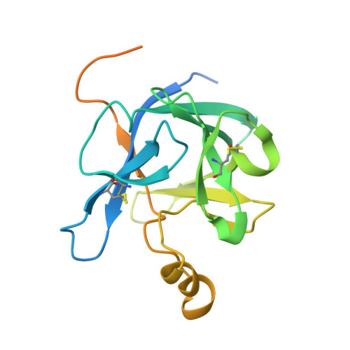
    Unique among fibroblast growth factors (FGFs), FGF19, -21, and -23 act in an endocrine fashion to regulate energy, bile acid, glucose, lipid, phosphate, and vitamin D homeostasis. These FGFs require the presence of Klotho/betaKlotho in their target tissues. Here, we present the crystal structures of FGF19 alone and FGF23 in complex with sucrose octasulfate, a disaccharide chemically related to heparin. The conformation of the heparin-binding region between beta strands 10 and 12 in FGF19 and FGF23 diverges completely from the common conformation adopted by paracrine-acting FGFs. A cleft between this region and the beta1-beta2 loop, the other heparin-binding region, precludes direct interaction between heparin/heparan sulfate and backbone atoms of FGF19/23. This reduces the heparin-binding affinity of these ligands and confers endocrine function. Klotho/betaKlotho have evolved as a compensatory mechanism for the poor ability of heparin/heparan sulfate to promote binding of FGF19, -21, and -23 to their cognate receptors.


  • Organizational Affiliation
    • Department of Pharmacology, New York University School of Medicine, New York, NY 10016, USA.

Macromolecule Content 

  • Total Structure Weight: 43.43 kDa 
  • Atom Count: 2,140 
  • Modeled Residue Count: 272 
  • Deposited Residue Count: 388 
  • Unique protein chains: 1

Macromolecules

Find similar proteins by:|  3D Structure
Entity ID: 1
MoleculeChains  Sequence LengthOrganismDetailsImage
Fibroblast growth factor 19
A, B
194Homo sapiensMutation(s): 0 
Gene Names: FGF19
UniProt & NIH Common Fund Data Resources
Find proteins for O95750 (Homo sapiens)
Explore O95750 
Go to UniProtKB:  O95750
PHAROS:  O95750
GTEx:  ENSG00000162344 
Entity Groups
Sequence Clusters30% Identity50% Identity70% Identity90% Identity95% Identity100% Identity
UniProt GroupO95750
Sequence Annotations
Expand
Reference Sequence

Experimental Data & Validation

Experimental Data

  • Method: X-RAY DIFFRACTION
  • Resolution: 1.80 Å
  • R-Value Free:  0.285 (Depositor), 0.284 (DCC) 
  • R-Value Work:  0.251 (Depositor), 0.249 (DCC) 
Space Group: P 3
Unit Cell:
Length ( Å )Angle ( ˚ )
a = 67.297α = 90
b = 67.297β = 90
c = 54.557γ = 120
Software Package:
Software NamePurpose
HKL-2000data collection
AMoREphasing
CNSrefinement
HKL-2000data reduction
HKL-2000data scaling

Structure Validation

View Full Validation Report


Entry History 

Deposition Data

Revision History  (Full details and data files)

  • Version 1.0: 2007-04-10
    Type: Initial release
  • Version 1.1: 2008-05-01
    Changes: Version format compliance
  • Version 1.2: 2011-07-13
    Changes: Version format compliance
  • Version 1.3: 2023-08-30
    Changes: Data collection, Database references, Refinement description
  • Version 1.4: 2024-10-16
    Changes: Structure summary