2OW3 | pdb_00002ow3

Glycogen synthase kinase-3 beta in complex with bis-(indole)maleimide pyridinophane inhibitor


Experimental Data Snapshot

  • Method: X-RAY DIFFRACTION
  • Resolution: 2.80 Å
  • R-Value Free: 
    0.295 (Depositor), 0.257 (DCC) 
  • R-Value Work: 
    0.246 (Depositor), 0.232 (DCC) 
  • R-Value Observed: 
    0.248 (Depositor) 

Starting Model: experimental
View more details

wwPDB Validation 3D Report Full Report

Validation slider image for 2OW3

Ligand Structure Quality Assessment 


This is version 1.5 of the entry. See complete history

Literature

Novel bis(indolyl)maleimide pyridinophanes that are potent, selective inhibitors of glycogen synthase kinase-3.

Zhang, H.C.Bonaga, L.V.Ye, H.Derian, C.K.Damiano, B.P.Maryanoff, B.E.

(2007) Bioorg Med Chem Lett 17: 2863-2868

  • DOI: https://doi.org/10.1016/j.bmcl.2007.02.059
  • Primary Citation Related Structures: 
    2OW3

  • PubMed Abstract: 

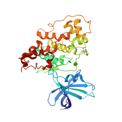
    Novel bis(indolyl)maleimide pyridinophanes 3, 9a, 9b, 10a, 10b, and 11 were prepared by cobalt-mediated [2+2+2] cycloaddition of an appropriate alpha,omega-diyne with an N,N-dialkylcyanamide. These macrocyclic heterophanes were found to be potent, selective inhibitors of glycogen synthase kinase-3beta. An X-ray structure of a co-crystal of GSK-3beta and 3 (IC(50)=3nM) depicts the hydrogen bonding and hydrophobic interactions in the ATP-binding pocket of this serine/threonine protein kinase.


  • Organizational Affiliation
    • Vascular Research Team, Johnson & Johnson Pharmaceutical Research & Development, Spring House, PA 19477-0776, USA. hzhang@prdus.jnj.com

Macromolecule Content 

  • Total Structure Weight: 81.25 kDa 
  • Atom Count: 5,681 
  • Modeled Residue Count: 699 
  • Deposited Residue Count: 704 
  • Unique protein chains: 1

Macromolecules

Find similar proteins by:|  3D Structure
Entity ID: 1
MoleculeChains  Sequence LengthOrganismDetailsImage
Glycogen synthase kinase-3 beta
A, B
352Homo sapiensMutation(s): 1 
Gene Names: GSK3B
EC: 2.7.11.26 (PDB Primary Data), 2.7.11.1 (UniProt)
UniProt & NIH Common Fund Data Resources
Find proteins for P49841 (Homo sapiens)
Explore P49841 
Go to UniProtKB:  P49841
PHAROS:  P49841
GTEx:  ENSG00000082701 
Entity Groups
Sequence Clusters30% Identity50% Identity70% Identity90% Identity95% Identity100% Identity
UniProt GroupP49841
Sequence Annotations
Expand
Reference Sequence

Small Molecules

Modified Residues  1 Unique
IDChains TypeFormula2D DiagramParent
PTR
Query on PTR
A, B
L-PEPTIDE LINKINGC9 H12 N O6 PTYR
Binding Affinity Annotations 
IDSourceBinding Affinity
BIM BindingDB:  2OW3 IC50: 3 (nM) from 1 assay(s)

Experimental Data & Validation

Experimental Data

  • Method: X-RAY DIFFRACTION
  • Resolution: 2.80 Å
  • R-Value Free:  0.295 (Depositor), 0.257 (DCC) 
  • R-Value Work:  0.246 (Depositor), 0.232 (DCC) 
  • R-Value Observed: 0.248 (Depositor) 
Space Group: P 1 21 1
Unit Cell:
Length ( Å )Angle ( ˚ )
a = 69.472α = 90
b = 117.329β = 103.22
c = 69.461γ = 90
Software Package:
Software NamePurpose
REFMACrefinement
MAR345data collection
MOSFLMdata reduction
SCALAdata scaling
CCP4phasing

Structure Validation

View Full Validation Report


Ligand Structure Quality Assessment 


Entry History 

Deposition Data

Revision History  (Full details and data files)

  • Version 1.0: 2008-02-19
    Type: Initial release
  • Version 1.1: 2011-07-13
    Changes: Version format compliance
  • Version 1.2: 2017-10-18
    Changes: Refinement description
  • Version 1.3: 2023-08-30
    Changes: Data collection, Database references, Derived calculations, Refinement description
  • Version 1.4: 2023-11-15
    Changes: Data collection
  • Version 1.5: 2024-11-13
    Changes: Structure summary