2OIH

Hepatitis Delta Virus gemonic ribozyme precursor with C75U mutation and bound to monovalent cation Tl+


Experimental Data Snapshot

  • Method: X-RAY DIFFRACTION
  • Resolution: 2.40 Å
  • R-Value Free: 0.288 
  • R-Value Work: 0.266 
  • R-Value Observed: 0.266 

wwPDB Validation   3D Report Full Report


This is version 1.3 of the entry. See complete history


Literature

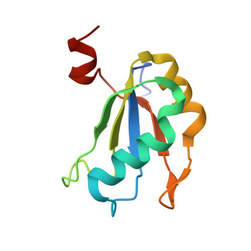
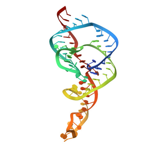
Structural roles of monovalent cations in the HDV ribozyme.

Ke, A.Ding, F.Batchelor, J.D.Doudna, J.A.

(2007) Structure 15: 281-287

  • DOI: https://doi.org/10.1016/j.str.2007.01.017
  • Primary Citation of Related Structures:  
    2OIH, 2OJ3

  • PubMed Abstract: 

    The hepatitis delta virus (HDV) ribozyme catalyzes viral RNA self-cleavage through general acid-base chemistry in which an active-site cytidine and at least one metal ion are involved. Monovalent metal ions support slow catalysis and were proposed to substitute for structural, but not catalytic, divalent metal ions in the RNA. To investigate the role of monovalent cations in ribozyme structure and function, we determined the crystal structure of the precursor HDV ribozyme in the presence of thallium ions (Tl(+)). Two Tl(+) ions can occupy a previously observed divalent metal ion hexahydrate-binding site located near the scissile phosphate, but are easily competed away by cobalt hexammine, a magnesium hexahydrate mimic and potent reaction inhibitor. Intriguingly, a third Tl(+) ion forms direct inner-sphere contacts with the ribose 2'-OH nucleophile and the pro-S(p) scissile phosphate oxygen. We discuss possible structural and catalytic implications of monovalent cation binding for the HDV ribozyme mechanism.


  • Organizational Affiliation

    Department of Molecular and Cell Biology, University of California at Berkeley, Berkeley, CA 94720, USA.


Macromolecules

Find similar proteins by:  (by identity cutoff)  |  3D Structure
Entity ID: 2
MoleculeChains Sequence LengthOrganismDetailsImage
U1 small nuclear ribonucleoprotein AB [auth A]100Homo sapiensMutation(s): 0 
Gene Names: SNRPA
UniProt & NIH Common Fund Data Resources
Find proteins for P09012 (Homo sapiens)
Explore P09012 
Go to UniProtKB:  P09012
PHAROS:  P09012
GTEx:  ENSG00000077312 
Entity Groups  
Sequence Clusters30% Identity50% Identity70% Identity90% Identity95% Identity100% Identity
UniProt GroupP09012
Sequence Annotations
Expand
  • Reference Sequence
Find similar nucleic acids by:  (by identity cutoff)  |  3D Structure
Entity ID: 1
MoleculeChains LengthOrganismImage
HDV ribozymeA [auth B]75N/A
Sequence Annotations
Expand
  • Reference Sequence
Small Molecules
Ligands 1 Unique
IDChains Name / Formula / InChI Key2D Diagram3D Interactions
TL
Query on TL

Download Ideal Coordinates CCD File 
C [auth B]
D [auth B]
E [auth B]
F [auth B]
G [auth B]
C [auth B],
D [auth B],
E [auth B],
F [auth B],
G [auth B],
H [auth B],
I [auth B],
J [auth B],
K [auth B],
L [auth B],
M [auth B],
N [auth B],
O [auth B],
P [auth B],
Q [auth B]
THALLIUM (I) ION
Tl
ZLUSCZLCHQSJRU-UHFFFAOYSA-N
Experimental Data & Validation

Experimental Data

  • Method: X-RAY DIFFRACTION
  • Resolution: 2.40 Å
  • R-Value Free: 0.288 
  • R-Value Work: 0.266 
  • R-Value Observed: 0.266 
  • Space Group: H 3 2
Unit Cell:
Length ( Å )Angle ( ˚ )
a = 107.962α = 90
b = 107.962β = 90
c = 189.119γ = 120
Software Package:
Software NamePurpose
CNSrefinement
HKL-2000data collection
HKL-2000data reduction
HKL-2000data scaling
AMoREphasing

Structure Validation

View Full Validation Report


Entry History 

Deposition Data

Revision History  (Full details and data files)

  • Version 1.0: 2007-03-27
    Type: Initial release
  • Version 1.1: 2008-05-01
    Changes: Version format compliance
  • Version 1.2: 2011-07-13
    Changes: Version format compliance
  • Version 1.3: 2023-12-27
    Changes: Data collection, Database references, Derived calculations