2OH4 | pdb_00002oh4

Crystal structure of Vegfr2 with a benzimidazole-urea inhibitor


Experimental Data Snapshot

  • Method: X-RAY DIFFRACTION
  • Resolution: 2.05 Å
  • R-Value Free: 
    0.231 (Depositor), 0.244 (DCC) 
  • R-Value Work: 
    0.187 (Depositor), 0.196 (DCC) 
  • R-Value Observed: 
    0.189 (Depositor) 

Starting Model: experimental
View more details

wwPDB Validation   3D Report Full Report


Ligand Structure Quality Assessment 


This is version 1.6 of the entry. See complete history


Literature

Discovery of Novel Benzimidazoles as Potent Inhibitors of TIE-2 and VEGFR-2 Tyrosine Kinase Receptors.

Hasegawa, M.Nishigaki, N.Washio, Y.Kano, K.Harris, P.A.Sato, H.Mori, I.West, R.I.Shibahara, M.Toyoda, H.Wang, L.Nolte, R.T.Veal, J.M.Cheung, M.

(2007) J Med Chem 50: 4453-4470

  • DOI: https://doi.org/10.1021/jm0611051
  • Primary Citation Related Structures: 
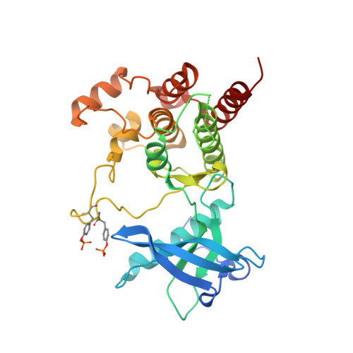
    2OH4

  • PubMed Abstract: 

    We herein disclose a novel chemical series of benzimidazole-ureas as inhibitors of VEGFR-2 and TIE-2 kinase receptors, both of which are implicated in angiogenesis. Structure-activity relationship (SAR) studies elucidated a critical role for the N1 nitrogen of both the benzimidazole (segment E) and urea (segment B) moieties. The SAR results were also supported by the X-ray crystallographic elucidation of the role of the N1 nitrogen and the urea moiety when the benzimidazole-urea compounds were bound to the VEGFR-2 enzyme. The left side phenyl ring (segment A) occupies the backpocket where a 3-hydrophobic substituent was favored for TIE-2 activity.


  • Organizational Affiliation
    • Tsukuba Research Laboratories, GlaxoSmithKline K.K., 43 Wadai, Tsukuba, Ibaraki 300-4247, Japan. masaichi.hasegawa@gsk.com

Macromolecules
Find similar proteins by:  (by identity cutoff)  |  3D Structure
Entity ID: 1
MoleculeChains Sequence LengthOrganismDetailsImage
Vascular endothelial growth factor receptor 2316Homo sapiensMutation(s): 0 
Gene Names: KDRFLK1VEGFR2
EC: 2.7.10.1
UniProt & NIH Common Fund Data Resources
Find proteins for P35968 (Homo sapiens)
Explore P35968 
Go to UniProtKB:  P35968
PHAROS:  P35968
GTEx:  ENSG00000128052 
Entity Groups  
Sequence Clusters30% Identity50% Identity70% Identity90% Identity95% Identity100% Identity
UniProt GroupP35968
Sequence Annotations
Expand
  • Reference Sequence
Small Molecules
Modified Residues  1 Unique
IDChains TypeFormula2D DiagramParent
PTR
Query on PTR
A
L-PEPTIDE LINKINGC9 H12 N O6 PTYR
Experimental Data & Validation

Experimental Data

  • Method: X-RAY DIFFRACTION
  • Resolution: 2.05 Å
  • R-Value Free:  0.231 (Depositor), 0.244 (DCC) 
  • R-Value Work:  0.187 (Depositor), 0.196 (DCC) 
  • R-Value Observed: 0.189 (Depositor) 
Space Group: P 21 21 21
Unit Cell:
Length ( Å )Angle ( ˚ )
a = 38.37α = 90
b = 94.25β = 90
c = 95.754γ = 90
Software Package:
Software NamePurpose
DENZOdata reduction
SCALEPACKdata scaling
REFMACrefinement
PDB_EXTRACTdata extraction
MAR345data collection
HKL-2000data reduction
AMoREphasing

Structure Validation

View Full Validation Report


Ligand Structure Quality Assessment 


Entry History 

Deposition Data

Revision History  (Full details and data files)

  • Version 1.0: 2007-09-18
    Type: Initial release
  • Version 1.1: 2011-07-13
    Changes: Advisory, Version format compliance
  • Version 1.2: 2017-06-14
    Changes: Advisory, Database references, Source and taxonomy, Structure summary
  • Version 1.3: 2017-10-18
    Changes: Refinement description
  • Version 1.4: 2023-08-30
    Changes: Data collection, Database references, Derived calculations, Refinement description
  • Version 1.5: 2023-11-15
    Changes: Data collection
  • Version 1.6: 2024-10-16
    Changes: Structure summary