2OE9 | pdb_00002oe9

High-pressure structure of pseudo-WT T4 Lysozyme


Experimental Data Snapshot

  • Method: X-RAY DIFFRACTION
  • Resolution: 2.01 Å
  • R-Value Free: 
    0.210 (Depositor), 0.166 (DCC) 
  • R-Value Work: 
    0.160 (Depositor), 0.163 (DCC) 
  • R-Value Observed: 
    0.163 (Depositor) 

Starting Model: experimental
View more details

wwPDB Validation 3D Report Full Report

Validation slider image for 2OE9

This is version 1.5 of the entry. See complete history

Literature

Structural Rigidity of a Large Cavity-containing Protein Revealed by High-pressure Crystallography.

Collins, M.D.Quillin, M.L.Hummer, G.Matthews, B.W.Gruner, S.M.

(2007) J Mol Biology 367: 752-763

  • DOI: https://doi.org/10.1016/j.jmb.2006.12.021
  • Primary Citation Related Structures: 
    2B6T, 2OE7, 2OE9, 2OEA

  • PubMed Abstract: 

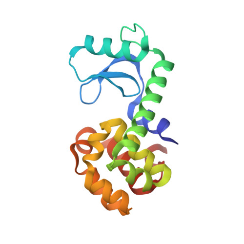
    Steric constraints, charged interactions and many other forces important to protein structure and function can be explored by mutagenic experiments. Research of this kind has led to a wealth of knowledge about what stabilizes proteins in their folded states. To gain a more complete picture requires that we perturb these structures in a continuous manner, something mutagenesis cannot achieve. With high pressure crystallographic methods it is now possible to explore the detailed properties of proteins while continuously varying thermodynamic parameters. Here, we detail the structural response of the cavity-containing mutant L99A of T4 lysozyme, as well as its pseudo wild-type (WT*) counterpart, to hydrostatic pressure. Surprisingly, the cavity has almost no effect on the pressure response: virtually the same changes are observed in WT* as in L99A under pressure. The cavity is most rigid, while other regions deform substantially. This implies that while some residues may increase the thermodynamic stability of a protein, they may also be structurally irrelevant. As recently shown, the cavity fills with water at pressures above 100 MPa while retaining its overall size. The resultant picture of the protein is one in which conformationally fluctuating side groups provide a liquid-like environment, but which also contribute to the rigidity of the peptide backbone.


  • Organizational Affiliation
    • Department of Physics, Cornell University, Ithaca, NY 14853, USA.

Macromolecule Content 

  • Total Structure Weight: 18.86 kDa 
  • Atom Count: 1,532 
  • Modeled Residue Count: 162 
  • Deposited Residue Count: 164 
  • Unique protein chains: 1

Macromolecules

Find similar proteins by:|  3D Structure
Entity ID: 1
MoleculeChains  Sequence LengthOrganismDetailsImage
LysozymeA [auth X]164Tequatrovirus T4Mutation(s): 2 
Gene Names: E
EC: 3.2.1.17
UniProt
Find proteins for P00720 (Enterobacteria phage T4)
Explore P00720 
Go to UniProtKB:  P00720
Entity Groups
Sequence Clusters30% Identity50% Identity70% Identity90% Identity95% Identity100% Identity
UniProt GroupP00720
Sequence Annotations
Expand
Reference Sequence

Experimental Data & Validation

Experimental Data

  • Method: X-RAY DIFFRACTION
  • Resolution: 2.01 Å
  • R-Value Free:  0.210 (Depositor), 0.166 (DCC) 
  • R-Value Work:  0.160 (Depositor), 0.163 (DCC) 
  • R-Value Observed: 0.163 (Depositor) 
Space Group: P 32 2 1
Unit Cell:
Length ( Å )Angle ( ˚ )
a = 60.554α = 90
b = 60.554β = 90
c = 95.567γ = 120
Software Package:
Software NamePurpose
DENZOdata reduction
SCALEPACKdata scaling
REFMACrefinement
PDB_EXTRACTdata extraction
ADSCdata collection

Structure Validation

View Full Validation Report


Entry History 

Deposition Data

Revision History  (Full details and data files)

  • Version 1.0: 2007-01-30
    Type: Initial release
  • Version 1.1: 2008-05-01
    Changes: Version format compliance
  • Version 1.2: 2011-07-13
    Changes: Version format compliance
  • Version 1.3: 2017-10-18
    Changes: Refinement description
  • Version 1.4: 2021-10-20
    Changes: Database references, Derived calculations
  • Version 1.5: 2023-08-30
    Changes: Data collection, Refinement description