2O7U

Crystal structure of K206E/K296E mutant of the N-terminal half molecule of human transferrin


Experimental Data Snapshot

  • Method: X-RAY DIFFRACTION
  • Resolution: 2.80 Å
  • R-Value Free: 0.259 
  • R-Value Work: 0.230 
  • R-Value Observed: 0.231 

wwPDB Validation   3D Report Full Report


This is version 1.5 of the entry. See complete history


Literature

Structures of two mutants that probe the role in iron release of the dilysine pair in the N-lobe of human transferrin.

Baker, H.M.Nurizzo, D.Mason, A.B.Baker, E.N.

(2007) Acta Crystallogr D Biol Crystallogr 63: 408-414

  • DOI: https://doi.org/10.1107/S0907444907000182
  • Primary Citation of Related Structures:  
    2O7U, 2O84

  • PubMed Abstract: 

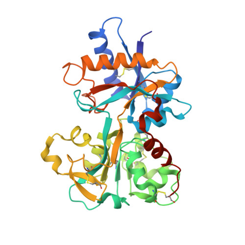
    Iron uptake by humans depends on the ability of the serum protein transferrin (Tf) to bind iron as Fe(3+) with high affinity but reversibly. Iron release into cells occurs through receptor-mediated endocytosis, aided by the lower endosomal pH of about 5.5. The protonation of a hydrogen-bonded pair of lysines, Lys206 and Lys296, adjacent to the N-lobe iron site of Tf has been proposed to create a repulsive interaction that stimulates domain opening and iron release. The crystal structures of two mutants, K206E (in which Lys206 is mutated to Glu) and K206E/K296E (in which both lysines are mutated to Glu), have been determined. The K206E structure (2.6 A resolution; R = 0.213, R(free) = 0.269) shows that a salt bridge is formed between Glu206 and Lys296, thus explaining the drastically slower iron release by this mutant. The K206E/K296E double-mutant structure (2.8 A resolution; R = 0.232, R(free) = 0.259) shows that the Glu296 side chain moves away from Glu206, easing any repulsive interaction and instead interacting with the iron ligand His249. The evident conformational flexibility is consistent with an alternative model for the operation of the dilysine pair in iron release in which it facilitates concerted proton transfer to the tyrosine ligand Tyr188 as one step in the weakening of iron binding.


  • Organizational Affiliation

    School of Biological Sciences, University of Auckland, Private Bag 92019, Auckland, New Zealand.


Macromolecules
Find similar proteins by:  (by identity cutoff)  |  3D Structure
Entity ID: 1
MoleculeChains Sequence LengthOrganismDetailsImage
Serotransferrin337Homo sapiensMutation(s): 2 
UniProt & NIH Common Fund Data Resources
Find proteins for P02787 (Homo sapiens)
Explore P02787 
Go to UniProtKB:  P02787
PHAROS:  P02787
GTEx:  ENSG00000091513 
Entity Groups  
Sequence Clusters30% Identity50% Identity70% Identity90% Identity95% Identity100% Identity
UniProt GroupP02787
Sequence Annotations
Expand
  • Reference Sequence
Small Molecules
Ligands 2 Unique
IDChains Name / Formula / InChI Key2D Diagram3D Interactions
CO3
Query on CO3

Download Ideal Coordinates CCD File 
AA [auth I]
K [auth B]
M [auth A]
O [auth C]
Q [auth D]
AA [auth I],
K [auth B],
M [auth A],
O [auth C],
Q [auth D],
S [auth E],
U [auth F],
W [auth G],
Y [auth H]
CARBONATE ION
C O3
BVKZGUZCCUSVTD-UHFFFAOYSA-L
FE
Query on FE

Download Ideal Coordinates CCD File 
J [auth B]
L [auth A]
N [auth C]
P [auth D]
R [auth E]
J [auth B],
L [auth A],
N [auth C],
P [auth D],
R [auth E],
T [auth F],
V [auth G],
X [auth H],
Z [auth I]
FE (III) ION
Fe
VTLYFUHAOXGGBS-UHFFFAOYSA-N
Experimental Data & Validation

Experimental Data

  • Method: X-RAY DIFFRACTION
  • Resolution: 2.80 Å
  • R-Value Free: 0.259 
  • R-Value Work: 0.230 
  • R-Value Observed: 0.231 
  • Space Group: C 1 2 1
Unit Cell:
Length ( Å )Angle ( ˚ )
a = 169.462α = 90
b = 97.897β = 90.01
c = 208.953γ = 90
Software Package:
Software NamePurpose
SCALAdata scaling
PHASERphasing
REFMACrefinement
PDB_EXTRACTdata extraction
MAR345data collection
MOSFLMdata reduction
CCP4data scaling

Structure Validation

View Full Validation Report


Entry History 

Deposition Data

Revision History  (Full details and data files)

  • Version 1.0: 2007-01-23
    Type: Initial release
  • Version 1.1: 2008-05-01
    Changes: Version format compliance
  • Version 1.2: 2011-07-13
    Changes: Advisory, Version format compliance
  • Version 1.3: 2017-10-18
    Changes: Refinement description
  • Version 1.4: 2021-10-20
    Changes: Database references, Derived calculations
  • Version 1.5: 2023-08-30
    Changes: Data collection, Refinement description