Evolution of the redox function in mammalian apurinic/apyrimidinic endonuclease
Georgiadis, M.M., Luo, M., Gaur, R.K., Delaplane, S., Li, X., Kelley, M.R.(2008) Mutat Res 643: 54-63
- PubMed: 18579163
- DOI: https://doi.org/10.1016/j.mrfmmm.2008.04.008
- Primary Citation of Related Structures:
2O3C, 2O3H - PubMed Abstract:
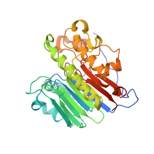
Human apurinic/apyrimidinic endonuclease (hApe1) encodes two important functional activities: an essential base excision repair (BER) activity and a redox activity that regulates expression of a number of genes through reduction of their transcription factors, AP-1, NFkappaB, HIF-1alpha, CREB, p53 and others. The BER function is highly conserved from prokaryotes (E. coli exonuclease III) to humans (hApe1). Here, we provide evidence supporting a redox function unique to mammalian Apes. An evolutionary analysis of Ape sequences reveals that, of the 7 Cys residues, Cys 93, 99, 208, 296, and 310 are conserved in both mammalian and non-mammalian vertebrate Apes, while Cys 65 is unique to mammalian Apes. In the zebrafish Ape (zApe), selected as the vertebrate sequence most distant from human, the residue equivalent to Cys 65 is Thr 58. The wild-type zApe enzyme was tested for redox activity in both in vitro EMSA and transactivation assays and found to be inactive, similar to C65A hApe1. Substitution of Thr 58 with Cys in zApe, however, resulted in a redox active enzyme, suggesting that a Cys residue in this position is indeed critical for redox function. In order to further probe differences between redox active and inactive enzymes, we have determined the crystal structures of vertebrate redox inactive enzymes, the C65A human Ape1 enzyme and the zApe enzyme at 1.9 and 2.3A, respectively. Our results provide new insights on the redox function and highlight a dramatic gain-of-function activity for Ape1 in mammals not found in non-mammalian vertebrates or lower organisms.
- Department of Biochemistry and Molecular Biology, Indiana University School of Medicine, 635 Barnhill Drive, Indianapolis, IN 46202-5122, United States. mgeorgia@iupui.edu
Organizational Affiliation:


