The Three-dimensional Structure of N-Succinyldiaminopimelate Aminotransferase from Mycobacterium tuberculosis
Weyand, S., Kefala, G., Weiss, M.S.(2007) J Mol Biology 367: 825-838
- PubMed: 17292400 Search on PubMed
- DOI: https://doi.org/10.1016/j.jmb.2007.01.023
- Primary Citation Related Structures:
2O0R - PubMed Abstract:
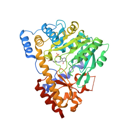
Inhibitors of the enzymes of the lysine biosynthetic pathway are considered promising lead compounds for the design of new antibacterial drugs, because the pathway appears to be indispensable for bacteria and because it is absent in humans. As part of our efforts to structurally characterize all enzymes of this pathway in Mycobacterium tuberculosis (Mtb), we have determined the three-dimensional structure of N-succinyldiaminopimelate aminotransferase (DapC, DAP-AT, Rv0858c) to a resolution of 2.0 A. This structure is the first DAP-AT structure reported to date. The orthorhombic crystals of Mtb-DAP-AT contain one functional dimer exhibiting C(2) symmetry in the asymmetric unit. The homodimer displays the typical S-shape of class I pyridoxal-5'-phosphate (PLP)-binding proteins. The two active sites of the dimer both feature an internal aldimine with the co-factor PLP covalently bound to the Lys232, although neither substrate nor co-factor had been added during protein production, purification and crystallization. Nine water molecules are conserved in the active site and form an intricate hydrogen-bonding network with the co-factor and the surrounding amino acid residues. Together with some residual difference electron density in the active site, this architecture permitted the building of external aldimine models of the enzyme with the substrates glutamate, the amine donor, and N-succinyl-2-amino-6-keto-pimelate, the amine acceptor. Based on these models, the amino acids relevant for substrate binding and specificity can be postulated. Furthermore, in the external aldimine model of N-succinyl-2-amino-6-keto-pimelate, the succinyl group overlaps with a glycerol binding site that has also been identified in both active sites of the Mtb-DAP-AT dimer. A comparison of the structure of Mtb-DAP-AT with other class I PLP-binding proteins, revealed that some inhibitors utilize the same binding site. Thus, the proposed models also provide an explanation for the mode of inhibition of Mtb-DAP-AT and they may be of help in the design of compounds, which are capable of inhibiting the enzyme. Last, but not least, a chloride binding helix exhibiting a peculiar amino acid sequence with a number of exposed hydrophobic side-chains was identified, which may be hypothesized as a putative docking site.
- EMBL Hamburg Outstation, DESY, Notkestr. 85, D-22603 Hamburg, Germany.
Organizational Affiliation: