2NZ2

Crystal structure of human argininosuccinate synthase in complex with aspartate and citrulline


Experimental Data Snapshot

  • Method: X-RAY DIFFRACTION
  • Resolution: 2.40 Å
  • R-Value Free: 0.265 
  • R-Value Work: 0.194 
  • R-Value Observed: 0.198 

wwPDB Validation   3D Report Full Report


Ligand Structure Quality Assessment 


This is version 1.3 of the entry. See complete history


Literature

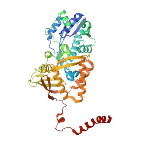
Structure of human argininosuccinate synthetase.

Karlberg, T.Collins, R.van den Berg, S.Flores, A.Hammarstrom, M.Hogbom, M.Holmberg Schiavone, L.Uppenberg, J.

(2008) Acta Crystallogr D Biol Crystallogr 64: 279-286

  • DOI: https://doi.org/10.1107/S0907444907067455
  • Primary Citation of Related Structures:  
    2NZ2

  • PubMed Abstract: 
  • Argininosuccinate synthetase catalyzes the transformation of citrulline and aspartate into argininosuccinate and pyrophosphate using the hydrolysis of ATP to AMP and pyrophosphate. This enzymatic process constitutes the rate-limiting step in both the urea and arginine-citrulline cycles ...

    Argininosuccinate synthetase catalyzes the transformation of citrulline and aspartate into argininosuccinate and pyrophosphate using the hydrolysis of ATP to AMP and pyrophosphate. This enzymatic process constitutes the rate-limiting step in both the urea and arginine-citrulline cycles. Previous studies have investigated the crystal structures of argininosuccinate synthetase from bacterial species. In this work, the first crystal structure of human argininosuccinate synthetase in complex with the substrates citrulline and aspartate is presented. The human enzyme is compared with structures of argininosuccinate synthetase from bacteria. In addition, the structure also provides new insights into the function of the numerous clinical mutations identified in patients with type I citrullinaemia (also known as classic citrullinaemia).


    Organizational Affiliation

    Structural Genomics Consortium, Karolinska Institutet, MBB/SGC, 171 77 Stockholm, Sweden.


Macromolecules
Find similar proteins by:  (by identity cutoff)  |  3D Structure
Entity ID: 1
MoleculeChainsSequence LengthOrganismDetailsImage
Argininosuccinate synthaseA413Homo sapiensMutation(s): 0 
Gene Names: ASS1ASS
EC: 6.3.4.5
UniProt & NIH Common Fund Data Resources
Find proteins for P00966 (Homo sapiens)
Explore P00966 
Go to UniProtKB:  P00966
PHAROS:  P00966
Entity Groups  
Sequence Clusters30% Identity50% Identity70% Identity90% Identity95% Identity100% Identity
UniProt GroupP00966
Protein Feature View
Expand
  • Reference Sequence
Small Molecules
Ligands 3 Unique
IDChainsName / Formula / InChI Key2D Diagram3D Interactions
CIR
Query on CIR

Download Ideal Coordinates CCD File 
D [auth A]CITRULLINE
C6 H13 N3 O3
RHGKLRLOHDJJDR-BYPYZUCNSA-N
 Ligand Interaction
ASP
Query on ASP

Download Ideal Coordinates CCD File 
C [auth A]ASPARTIC ACID
C4 H7 N O4
CKLJMWTZIZZHCS-REOHCLBHSA-N
 Ligand Interaction
NA
Query on NA

Download Ideal Coordinates CCD File 
B [auth A]SODIUM ION
Na
FKNQFGJONOIPTF-UHFFFAOYSA-N
 Ligand Interaction
Experimental Data & Validation

Experimental Data

  • Method: X-RAY DIFFRACTION
  • Resolution: 2.40 Å
  • R-Value Free: 0.265 
  • R-Value Work: 0.194 
  • R-Value Observed: 0.198 
  • Space Group: F 2 2 2
Unit Cell:
Length ( Å )Angle ( ˚ )
a = 95.94α = 90
b = 117.47β = 90
c = 155.15γ = 90
Software Package:
Software NamePurpose
REFMACrefinement
MAR345data collection
XDSdata reduction
XSCALEdata scaling
MOLREPphasing

Structure Validation

View Full Validation Report


Ligand Structure Quality Assessment 


Entry History 

Revision History  (Full details and data files)

  • Version 1.0: 2006-12-05
    Type: Initial release
  • Version 1.1: 2008-02-20
    Changes: Version format compliance
  • Version 1.2: 2011-07-13
    Changes: Derived calculations, Version format compliance
  • Version 1.3: 2017-10-18
    Changes: Refinement description