2NUN

The structure of the type III effector AvrB complexed with ADP


Experimental Data Snapshot

  • Method: X-RAY DIFFRACTION
  • Resolution: 2.40 Å
  • R-Value Free: 0.254 
  • R-Value Work: 0.232 
  • R-Value Observed: 0.232 

wwPDB Validation   3D Report Full Report


Ligand Structure Quality Assessment 


This is version 1.3 of the entry. See complete history


Literature

Type III Effector Activation via Nucleotide Binding, Phosphorylation, and Host Target Interaction.

Desveaux, D.Singer, A.U.Wu, A.J.McNulty, B.C.Musselwhite, L.Nimchuk, Z.Sondek, J.Dangl, J.L.

(2007) PLoS Pathog 3: e48

  • DOI: https://doi.org/10.1371/journal.ppat.0030048
  • Primary Citation of Related Structures:  
    2NUD, 2NUN

  • PubMed Abstract: 

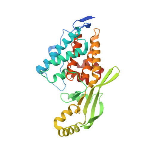
    The Pseudomonas syringae type III effector protein avirulence protein B (AvrB) is delivered into plant cells, where it targets the Arabidopsis RIN4 protein (resistance to Pseudomonas maculicula protein 1 [RPM1]-interacting protein). RIN4 is a regulator of basal host defense responses. Targeting of RIN4 by AvrB is recognized by the host RPM1 nucleotide-binding leucine-rich repeat disease resistance protein, leading to accelerated defense responses, cessation of pathogen growth, and hypersensitive host cell death at the infection site. We determined the structure of AvrB complexed with an AvrB-binding fragment of RIN4 at 2.3 A resolution. We also determined the structure of AvrB in complex with adenosine diphosphate bound in a binding pocket adjacent to the RIN4 binding domain. AvrB residues important for RIN4 interaction are required for full RPM1 activation. AvrB residues that contact adenosine diphosphate are also required for initiation of RPM1 function. Nucleotide-binding residues of AvrB are also required for its phosphorylation by an unknown Arabidopsis protein(s). We conclude that AvrB is activated inside the host cell by nucleotide binding and subsequent phosphorylation and, independently, interacts with RIN4. Our data suggest that activated AvrB, bound to RIN4, is indirectly recognized by RPM1 to initiate plant immune system function.


  • Organizational Affiliation

    Department of Biology, University of North Carolina at Chapel Hill, Chapel Hill, North Carolina, United States of America.


Macromolecules
Find similar proteins by:  (by identity cutoff)  |  3D Structure
Entity ID: 1
MoleculeChains Sequence LengthOrganismDetailsImage
Avirulence B protein323Pseudomonas savastanoi pv. glycineaMutation(s): 0 
Gene Names: avrB
UniProt
Find proteins for P13835 (Pseudomonas savastanoi pv. glycinea)
Explore P13835 
Go to UniProtKB:  P13835
Entity Groups  
Sequence Clusters30% Identity50% Identity70% Identity90% Identity95% Identity100% Identity
UniProt GroupP13835
Sequence Annotations
Expand
  • Reference Sequence
Small Molecules
Ligands 2 Unique
IDChains Name / Formula / InChI Key2D Diagram3D Interactions
ADP
Query on ADP

Download Ideal Coordinates CCD File 
B [auth A]ADENOSINE-5'-DIPHOSPHATE
C10 H15 N5 O10 P2
XTWYTFMLZFPYCI-KQYNXXCUSA-N
TRS
Query on TRS

Download Ideal Coordinates CCD File 
C [auth A]2-AMINO-2-HYDROXYMETHYL-PROPANE-1,3-DIOL
C4 H12 N O3
LENZDBCJOHFCAS-UHFFFAOYSA-O
Experimental Data & Validation

Experimental Data

  • Method: X-RAY DIFFRACTION
  • Resolution: 2.40 Å
  • R-Value Free: 0.254 
  • R-Value Work: 0.232 
  • R-Value Observed: 0.232 
  • Space Group: P 65
Unit Cell:
Length ( Å )Angle ( ˚ )
a = 122.717α = 90
b = 122.717β = 90
c = 64.091γ = 120
Software Package:
Software NamePurpose
CNSrefinement
DENZOdata reduction
SCALEPACKdata scaling
AMoREphasing

Structure Validation

View Full Validation Report


Ligand Structure Quality Assessment 


Entry History 

Deposition Data

Revision History  (Full details and data files)

  • Version 1.0: 2007-05-22
    Type: Initial release
  • Version 1.1: 2008-05-01
    Changes: Version format compliance
  • Version 1.2: 2011-07-13
    Changes: Version format compliance
  • Version 1.3: 2023-08-30
    Changes: Data collection, Database references, Derived calculations, Refinement description