2NSI | pdb_00002nsi

HUMAN INDUCIBLE NITRIC OXIDE SYNTHASE, ZN-FREE, SEITU COMPLEX


Experimental Data Snapshot

  • Method: X-RAY DIFFRACTION
  • Resolution: 3.00 Å
  • R-Value Free: 
    0.238 (Depositor) 
  • R-Value Work: 
    0.214 (Depositor) 
  • R-Value Observed: 
    0.214 (Depositor) 

wwPDB Validation 3D Report Full Report

Validation slider image for 2NSI

This is version 1.4 of the entry. See complete history

Literature

Crystal structures of zinc-free and -bound heme domain of human inducible nitric-oxide synthase. Implications for dimer stability and comparison with endothelial nitric-oxide synthase.

Li, H.Raman, C.S.Glaser, C.B.Blasko, E.Young, T.A.Parkinson, J.F.Whitlow, M.Poulos, T.L.

(1999) J Biological Chem 274: 21276-21284

  • DOI: https://doi.org/10.1074/jbc.274.30.21276
  • Primary Citation Related Structures: 
    1NSI, 2NSI

  • PubMed Abstract: 

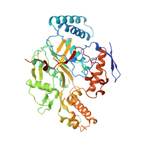
    The crystal structures of the heme domain of human inducible nitric-oxide synthase (NOS-2) in zinc-free and -bound states have been solved. In the zinc-free structure, two symmetry-related cysteine residues form a disulfide bond. In the zinc-bound state, these same two cysteine residues form part of a zinc-tetrathiolate (ZnS(4)) center indistinguishable from that observed in the endothelial isoform (NOS-3). As in NOS-3, ZnS(4) plays a key role in stabilizing intersubunit contacts and in maintaining the integrity of the cofactor (tetrahydrobiopterin) binding site of NOS-2. A comparison of NOS-2 and NOS-3 structures illustrates the conservation of quaternary structure, tertiary topology, and substrate and cofactor binding sites, in addition to providing insights on isoform-specific inhibitor design. The structural comparison also reveals that pterin binding does not preferentially stabilize the dimer interface of NOS-2 over NOS-3.


  • Organizational Affiliation
    • Department of Molecular Biology and Biochemistry, University of California at Irvine, Irvine, California 92697, USA.

Macromolecule Content 

  • Total Structure Weight: 203.53 kDa 
  • Atom Count: 13,985 
  • Modeled Residue Count: 1,680 
  • Deposited Residue Count: 1,724 
  • Unique protein chains: 1

Macromolecules

Find similar proteins by:|  3D Structure
Entity ID: 1
MoleculeChains  Sequence LengthOrganismDetailsImage
PROTEIN (NITRIC OXIDE SYNTHASE)
A, B, C, D
431Homo sapiensMutation(s): 0 
EC: 1.14.13.39
UniProt & NIH Common Fund Data Resources
Find proteins for P35228 (Homo sapiens)
Explore P35228 
Go to UniProtKB:  P35228
PHAROS:  P35228
GTEx:  ENSG00000007171 
Entity Groups
Sequence Clusters30% Identity50% Identity70% Identity90% Identity95% Identity100% Identity
UniProt GroupP35228
Sequence Annotations
Expand
Reference Sequence

Small Molecules

Ligands 4 Unique
IDChains Name / Formula / InChI Key2D Diagram3D Interactions
HEM

Query on HEM


Download:Ideal Coordinates CCD File
H [auth A],
M [auth B],
R [auth C],
W [auth D]
PROTOPORPHYRIN IX CONTAINING FE
C34 H32 Fe N4 O4
KABFMIBPWCXCRK-RGGAHWMASA-L
H4B

Query on H4B


Download:Ideal Coordinates CCD File
I [auth A],
N [auth B],
S [auth C],
X [auth D]
5,6,7,8-TETRAHYDROBIOPTERIN
C9 H15 N5 O3
FNKQXYHWGSIFBK-RPDRRWSUSA-N
ITU

Query on ITU


Download:Ideal Coordinates CCD File
J [auth A],
O [auth B],
T [auth C],
Y [auth D]
ETHYLISOTHIOUREA
C3 H8 N2 S
VFIZBHJTOHUOEK-UHFFFAOYSA-N
SO4

Query on SO4


Download:Ideal Coordinates CCD File
E [auth A]
F [auth A]
G [auth A]
K [auth B]
L [auth B]
E [auth A],
F [auth A],
G [auth A],
K [auth B],
L [auth B],
P [auth C],
Q [auth C],
U [auth D],
V [auth D]
SULFATE ION
O4 S
QAOWNCQODCNURD-UHFFFAOYSA-L
Binding Affinity Annotations 
IDSourceBinding Affinity
ITU BindingDB:  2NSI Ki: min: 5.2, max: 190 (nM) from 2 assay(s)
IC50: 160 (nM) from 1 assay(s)

Experimental Data & Validation

Experimental Data

  • Method: X-RAY DIFFRACTION
  • Resolution: 3.00 Å
  • R-Value Free:  0.238 (Depositor) 
  • R-Value Work:  0.214 (Depositor) 
  • R-Value Observed: 0.214 (Depositor) 
Space Group: P 43 21 2
Unit Cell:
Length ( Å )Angle ( ˚ )
a = 188.37α = 90
b = 188.37β = 90
c = 230.16γ = 90
Software Package:
Software NamePurpose
DENZOdata reduction
SCALEPACKdata scaling
X-PLORmodel building
X-PLORrefinement
X-PLORphasing

Structure Validation

View Full Validation Report


Entry History 

Deposition Data

Revision History  (Full details and data files)

  • Version 1.0: 2000-01-07
    Type: Initial release
  • Version 1.1: 2008-04-27
    Changes: Version format compliance
  • Version 1.2: 2011-07-13
    Changes: Version format compliance
  • Version 1.3: 2023-12-27
    Changes: Data collection, Database references, Derived calculations
  • Version 1.4: 2024-10-30
    Changes: Structure summary