2NR9

Crystal structure of GlpG, Rhomboid Peptidase from Haemophilus influenzae


Experimental Data Snapshot

  • Method: X-RAY DIFFRACTION
  • Resolution: 2.20 Å
  • R-Value Free: 0.295 
  • R-Value Work: 0.233 
  • R-Value Observed: 0.236 

wwPDB Validation   3D Report Full Report


This is version 1.4 of the entry. See complete history


Literature

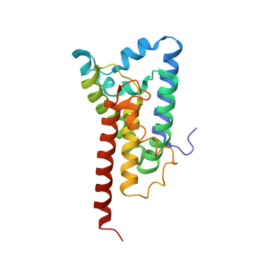
The crystal structure of the rhomboid peptidase from Haemophilus influenzae provides insight into intramembrane proteolysis.

Lemieux, M.J.Fischer, S.J.Cherney, M.M.Bateman, K.S.James, M.N.G.

(2007) Proc Natl Acad Sci U S A 104: 750-754

  • DOI: https://doi.org/10.1073/pnas.0609981104
  • Primary Citation of Related Structures:  
    2NR9

  • PubMed Abstract: 

    Rhomboid peptidases are members of a family of regulated intramembrane peptidases that cleave the transmembrane segments of integral membrane proteins. Rhomboid peptidases have been shown to play a major role in developmental processes in Drosophila and in mitochondrial maintenance in yeast. Most recently, the function of rhomboid peptidases has been directly linked to apoptosis. We have solved the structure of the rhomboid peptidase from Haemophilus influenzae (hiGlpG) to 2.2-A resolution. The phasing for the crystals of hiGlpG was provided mainly by molecular replacement, by using the coordinates of the Escherichia coli rhomboid (ecGlpG). The structural results on these rhomboid peptidases have allowed us to speculate on the catalytic mechanism of substrate cleavage in a membranous environment. We have identified the relative disposition of the nucleophilic serine to the general base/acid function of the conserved histidine. Modeling a tetrapeptide substrate in the context of the rhomboid structure reveals an oxyanion hole comprising the side chain of a second conserved histidine and the main-chain NH of the nucleophilic serine residue. In both hiGlpG and ecGlpG structures, a water molecule occupies this oxyanion hole.


  • Organizational Affiliation

    Group in Protein Structure and Function, Department of Biochemistry, University of Alberta, Edmonton, AB, Canada T6G 2H7.


Macromolecules
Find similar proteins by:  (by identity cutoff)  |  3D Structure
Entity ID: 1
MoleculeChains Sequence LengthOrganismDetailsImage
Protein glpG homolog196Haemophilus influenzae 86-028NPMutation(s): 2 
Gene Names: glpG
EC: 3.4.21.105
Membrane Entity: Yes 
UniProt
Find proteins for P44783 (Haemophilus influenzae (strain ATCC 51907 / DSM 11121 / KW20 / Rd))
Explore P44783 
Go to UniProtKB:  P44783
Entity Groups  
Sequence Clusters30% Identity50% Identity70% Identity90% Identity95% Identity100% Identity
UniProt GroupP44783
Sequence Annotations
Expand
  • Reference Sequence
Experimental Data & Validation

Experimental Data

  • Method: X-RAY DIFFRACTION
  • Resolution: 2.20 Å
  • R-Value Free: 0.295 
  • R-Value Work: 0.233 
  • R-Value Observed: 0.236 
  • Space Group: C 1 2 1
Unit Cell:
Length ( Å )Angle ( ˚ )
a = 121.288α = 90
b = 35.047β = 104.02
c = 52.931γ = 90
Software Package:
Software NamePurpose
REFMACrefinement
Blu-Icedata collection
DENZOdata reduction
SCALEPACKdata scaling
MOLREPphasing

Structure Validation

View Full Validation Report


Entry History 

Deposition Data

Revision History  (Full details and data files)

  • Version 1.0: 2006-11-14
    Type: Initial release
  • Version 1.1: 2008-05-01
    Changes: Version format compliance
  • Version 1.2: 2011-07-13
    Changes: Advisory, Derived calculations, Source and taxonomy, Version format compliance
  • Version 1.3: 2021-10-20
    Changes: Database references, Derived calculations
  • Version 1.4: 2023-08-30
    Changes: Data collection, Refinement description