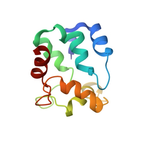
Solution structure of Ca2+-free rat beta-parvalbumin (oncomodulin).
Henzl, M.T., Tanner, J.J.(2007) Protein Sci 16: 1914-1926
- PubMed: 17766386 Search on PubMedSearch on PubMed Central
- DOI: https://doi.org/10.1110/ps.072837307
- Primary Citation Related Structures:
2NLN - PubMed Abstract:
Relative to other parvalbumin isoforms, the mammalian beta-parvalbumin (oncomodulin) displays attenuated divalent ion affinity. High-resolution structural data for the Ca(2+)-bound protein have provided little insight into the physical basis for this behavior, prompting an examination of the unliganded state. This article describes the solution structure and peptide backbone dynamics of Ca(2+)-free rat beta-parvalbumin (beta-PV). Ca(2+) removal evidently provokes significant structural alterations. Interaction between the D helix and the AB domain in the Ca(2+)-bound protein is greatly diminished in the apo-form, permitting the D helix to straighten. There is also a significant reorganization of the hydrophobic core and a concomitant remodeling of the interface between the AB and CD-EF domains. These modifications perturb the orientation of the C and D helices, and the energetic penalty associated with their reversal could contribute to the low-affinity signature of the CD site. By contrast, Ca(2+) removal causes a comparatively minor perturbation of the E and F helices, consistent with the more typical divalent ion affinity observed for the EF site. Ca(2+)-free rat beta-PV retains structural rigidity on the picosecond-nanosecond timescale. At 20 degrees C, the majority of amide vectors show no evidence for motion on timescales above 20 ps, and the average order parameter for the entire molecule is 0.92.
- Department of Biochemistry, University of Missouri-Columbia, Columbia, Missouri 65211, USA. henzlm@missouri.edu
Organizational Affiliation: