Membrane Interaction of the Glycosyltransferase WaaG.
Liebau, J., Pettersson, P., Szpryngiel, S., Maler, L.(2015) Biophys J 109: 552-563
- PubMed: 26244737
- DOI: https://doi.org/10.1016/j.bpj.2015.06.036
- Primary Citation of Related Structures:
2N58 - PubMed Abstract:
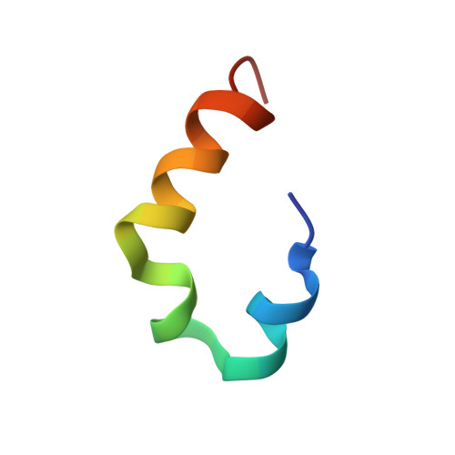
The glycosyltransferase WaaG is involved in the synthesis of lipopolysaccharides that constitute the outer leaflet of the outer membrane in Gram-negative bacteria such as Escherichia coli. WaaG has been identified as a potential antibiotic target, and inhibitor scaffolds have previously been investigated. WaaG is located at the cytosolic side of the inner membrane, where the enzyme catalyzes the transfer of the first outer-core glucose to the inner core of nascent lipopolysaccharides. Here, we characterized the binding of WaaG to membrane models designed to mimic the inner membrane of E. coli. Based on the crystal structure, we identified an exposed and largely α-helical 30-residue sequence, with a net positive charge and several aromatic amino acids, as a putative membrane-interacting region of WaaG (MIR-WaaG). We studied the peptide corresponding to this sequence, along with its bilayer interactions, using circular dichroism, fluorescence quenching, fluorescence anisotropy, and NMR. In the presence of dodecylphosphocholine, MIR-WaaG was observed to adopt a three-dimensional structure remarkably similar to the segment in the crystal structure. We found that the membrane interaction of WaaG is conferred at least in part by MIR-WaaG and that electrostatic interactions play a key role in binding. Moreover, we propose a mechanism of anchoring WaaG to the inner membrane of E. coli, where the central part of MIR-WaaG inserts into one leaflet of the bilayer. In this model, electrostatic interactions as well as surface-exposed Tyr residues bind WaaG to the membrane.
- Department of Biochemistry and Biophysics, Stockholm University, Stockholm, Sweden.
Organizational Affiliation: