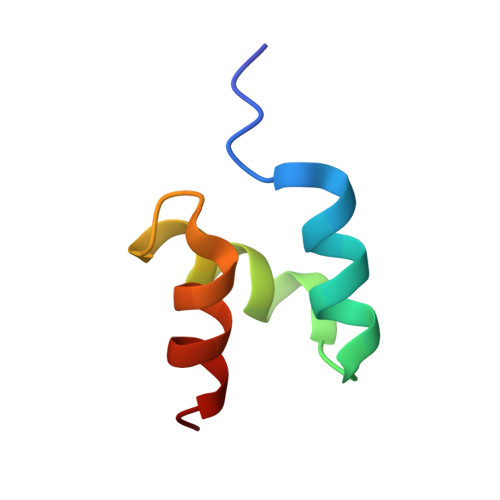
2MRO

Structure of the complex of ubiquitin and the UBA domain from DNA-damage-inducible 1 protein (Ddi1)


Experimental Data Snapshot

  • Method: SOLUTION NMR
  • Conformers Calculated: 10 
  • Conformers Submitted: 10 
  • Selection Criteria: structures with the least restraint violations 

wwPDB Validation   3D Report Full Report


This is version 1.4 of the entry. See complete history


Literature

DNA-Damage-Inducible 1 Protein (Ddi1) Contains an Uncharacteristic Ubiquitin-like Domain that Binds Ubiquitin.

Nowicka, U.Zhang, D.Walker, O.Krutauz, D.Castaneda, C.A.Chaturvedi, A.Chen, T.Y.Reis, N.Glickman, M.H.Fushman, D.

(2015) Structure 23: 542-557

  • DOI: https://doi.org/10.1016/j.str.2015.01.010
  • Primary Citation of Related Structures:  
    2MR9, 2MRO, 2MRP, 2MWS

  • PubMed Abstract: 

    Ddi1 belongs to a family of shuttle proteins targeting polyubiquitinated substrates for proteasomal degradation. Unlike the other proteasomal shuttles, Rad23 and Dsk2, Ddi1 remains an enigma: its function is not fully understood and structural properties are poorly characterized. We determined the structure and binding properties of the ubiquitin-like (UBL) and ubiquitin-associated (UBA) domains of Ddi1 from Saccharomyces cerevisiae. We found that while Ddi1UBA forms a characteristic UBA:ubiquitin complex, Ddi1UBL has entirely uncharacteristic binding preferences. Despite having a ubiquitin-like fold, Ddi1UBL does not interact with typical UBL receptors but unexpectedly binds ubiquitin, forming a unique interface mediated by hydrophobic contacts and by salt bridges between oppositely charged residues of Ddi1UBL and ubiquitin. In stark contrast to ubiquitin and other UBLs, the β-sheet surface of Ddi1UBL is negatively charged and therefore is recognized in a completely different way. The dual functionality of Ddi1UBL, capable of binding both ubiquitin and proteasome, suggests an intriguing mechanism for Ddi1 as a proteasomal shuttle.


  • Organizational Affiliation

    Department of Chemistry and Biochemistry, Center for Biomolecular Structure and Organization, University of Maryland, College Park, MD 20742, USA.


Macromolecules
Find similar proteins by:  (by identity cutoff)  |  3D Structure
Entity ID: 1
MoleculeChains Sequence LengthOrganismDetailsImage
Polyubiquitin-B76Homo sapiensMutation(s): 0 
Gene Names: UBB
UniProt & NIH Common Fund Data Resources
Find proteins for P0CG47 (Homo sapiens)
Explore P0CG47 
Go to UniProtKB:  P0CG47
PHAROS:  P0CG47
GTEx:  ENSG00000170315 
Entity Groups  
Sequence Clusters30% Identity50% Identity70% Identity90% Identity95% Identity100% Identity
UniProt GroupP0CG47
Sequence Annotations
Expand
  • Reference Sequence
Find similar proteins by:  (by identity cutoff)  |  3D Structure
Entity ID: 2
MoleculeChains Sequence LengthOrganismDetailsImage
DNA damage-inducible protein 144Saccharomyces cerevisiae S288CMutation(s): 0 
Gene Names: DDI1VSM1YER143W
UniProt
Find proteins for P40087 (Saccharomyces cerevisiae (strain ATCC 204508 / S288c))
Explore P40087 
Go to UniProtKB:  P40087
Entity Groups  
Sequence Clusters30% Identity50% Identity70% Identity90% Identity95% Identity100% Identity
UniProt GroupP40087
Sequence Annotations
Expand
  • Reference Sequence
Experimental Data & Validation

Experimental Data

  • Method: SOLUTION NMR
  • Conformers Calculated: 10 
  • Conformers Submitted: 10 
  • Selection Criteria: structures with the least restraint violations 

Structure Validation

View Full Validation Report


Entry History 

Deposition Data

Revision History  (Full details and data files)

  • Version 1.0: 2015-02-04
    Type: Initial release
  • Version 1.1: 2015-03-18
    Changes: Database references
  • Version 1.2: 2019-11-06
    Changes: Data collection, Database references, Experimental preparation
  • Version 1.3: 2023-06-14
    Changes: Database references, Other
  • Version 1.4: 2024-05-22
    Changes: Data collection