2MOL | pdb_00002mol

3D NMR structure of the cytoplasmic rhodanese domain of the full-length inner membrane protein YgaP from Escherichia coli


Experimental Data Snapshot

  • Method: SOLUTION NMR
  • Conformers Calculated: 100 
  • Conformers Submitted: 10 
  • Selection Criteria: target function 

wwPDB Validation 3D Report Full Report

Validation slider image for 2MOL

This is version 1.4 of the entry. See complete history

Literature

Solution NMR Structure and Functional Analysis of the Integral Membrane Protein YgaP from Escherichia coli.

Eichmann, C.Tzitzilonis, C.Bordignon, E.Maslennikov, I.Choe, S.Riek, R.

(2014) J Biological Chem 289: 23482-23503

  • DOI: https://doi.org/10.1074/jbc.M114.571935
  • Primary Citation Related Structures: 
    2MOI, 2MOL, 2MPN

  • PubMed Abstract: 

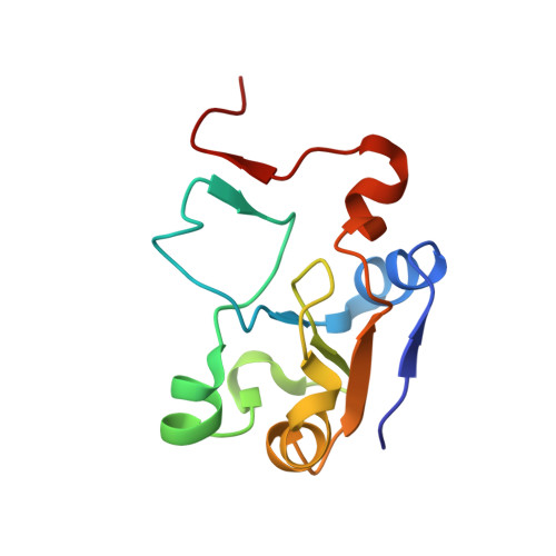
    The solution NMR structure of the α-helical integral membrane protein YgaP from Escherichia coli in mixed 1,2-diheptanoyl-sn-glycerol-3-phosphocholine/1-myristoyl-2-hydroxy-sn-glycero-3-phospho-(1'-rac-glycerol) micelles is presented. In these micelles, YgaP forms a homodimer with the two transmembrane helices being the dimer interface, whereas the N-terminal cytoplasmic domain includes a rhodanese-fold in accordance to its sequence homology to the rhodanese family of sulfurtransferases. The enzymatic sulfur transfer activity of full-length YgaP as well as of the N-terminal rhodanese domain only was investigated performing a series of titrations with sodium thiosulfate and potassium cyanide monitored by NMR and EPR. The data indicate the thiosulfate concentration-dependent addition of several sulfur atoms to the catalytic Cys-63, which process can be reversed by the addition of potassium cyanide. The catalytic reaction induces thereby conformational changes within the rhodanese domain, as well as on the transmembrane α-helices of YgaP. These results provide insights into a potential mechanism of YgaP during the catalytic thiosulfate activity in vivo.


  • Organizational Affiliation
    • From the Laboratory of Physical Chemistry, Swiss Federal Institute of Technology, ETH-Hönggerberg, CH-8093 Zürich, Switzerland and.

Macromolecule Content 

  • Total Structure Weight: 11.72 kDa 
  • Atom Count: 824 
  • Modeled Residue Count: 108 
  • Deposited Residue Count: 108 
  • Unique protein chains: 1

Macromolecules

Find similar proteins by:|  3D Structure
Entity ID: 1
MoleculeChains  Sequence LengthOrganismDetailsImage
Inner membrane protein YgaP108Escherichia coli K-12Mutation(s): 0 
Gene Names: b2668JW2643ygaPygaP b2668 JW2643
EC: 2.8.1.1
Membrane Entity: Yes 
UniProt
Find proteins for P55734 (Escherichia coli (strain K12))
Explore P55734 
Go to UniProtKB:  P55734
Entity Groups
Sequence Clusters30% Identity50% Identity70% Identity90% Identity95% Identity100% Identity
UniProt GroupP55734
Sequence Annotations
Expand
Reference Sequence

Experimental Data & Validation

Experimental Data

  • Method: SOLUTION NMR
  • Conformers Calculated: 100 
  • Conformers Submitted: 10 
  • Selection Criteria: target function 

Structure Validation

View Full Validation Report


Entry History 

Deposition Data

Revision History  (Full details and data files)

  • Version 1.0: 2014-06-25
    Type: Initial release
  • Version 1.1: 2014-08-13
    Changes: Database references
  • Version 1.2: 2014-09-17
    Changes: Database references
  • Version 1.3: 2023-06-14
    Changes: Data collection, Database references, Other
  • Version 1.4: 2024-05-15
    Changes: Data collection, Database references