2MHA | pdb_00002mha

CRYSTAL STRUCTURE OF THE MAJOR HISTOCOMPATIBILITY COMPLEX CLASS I H-2KB MOLECULE CONTAINING A SINGLE VIRAL PEPTIDE: IMPLICATIONS FOR PEPTIDE BINDING AND T-CELL RECEPTOR RECOGNITION


Experimental Data Snapshot

  • Method: X-RAY DIFFRACTION
  • Resolution: 2.50 Å
  • R-Value Work: 
    0.184 (Depositor) 
  • R-Value Observed: 
    0.184 (Depositor) 

wwPDB Validation 3D Report Full Report

Validation slider image for 2MHA

This is version 1.5 of the entry. See complete history

Literature

Crystal structure of the major histocompatibility complex class I H-2Kb molecule containing a single viral peptide: implications for peptide binding and T-cell receptor recognition.

Zhang, W.Young, A.C.Imarai, M.Nathenson, S.G.Sacchettini, J.C.

(1992) Proc Natl Acad Sci U S A 89: 8403-8407

  • DOI: https://doi.org/10.1073/pnas.89.17.8403
  • Primary Citation Related Structures: 
    2MHA

  • PubMed Abstract: 

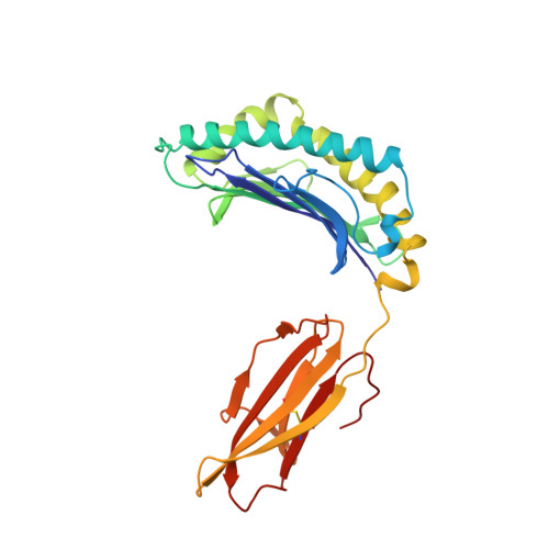
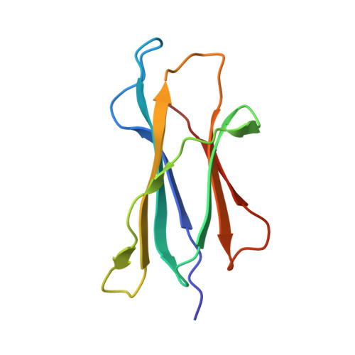
    To study the structure of a homogenous major histocompatibility complex (MHC) class I molecule containing a single bound peptide, a complex of recombinant mouse H-2Kb, beta 2-microglobulin (beta 2m), and a fragment of the vesicular stomatitis virus (VSV) nuclear capsid protein, VSV-(N52-59) octapeptide (Arg-Gly-Tyr-Val-Tyr-Gln-Gly-Leu), was prepared by exploiting a high-yield bacterial expression system and in vitro cocomplex formation. The structure of mouse H-2Kb revealed its similarity to three human class I HLA molecules, consistent with the high primary sequence homology and common function of these peptide-presenting molecules. Electron density was located in the peptide-binding groove, to which a single peptide in a unique conformation was unambiguously fit. The peptide extends the length of the groove, parallel to the alpha-helices, and assumes an extended, mostly beta-strand conformation. The peptide is constrained within the groove by hydrogen bonding of its main-chain atoms and by contacts of its side chains with the H-2Kb molecule. The amino-terminal nitrogen atom of the peptide forms a hydrogen bond with the hydroxyl group of Tyr-171 of H-2Kb at one end of the groove, while the carboxyl-terminal oxygen forms a hydrogen bond with the hydroxyl group of Tyr-84 at the other end. Since the amino acids at both ends are conserved among human and mouse MHC molecules, this anchoring of each end of the peptide appears to be a general feature of peptide-MHC class I molecule binding and imposes restrictions on its length. The side chains of residues Tyr-3, Tyr-5, and Leu-8 of the VSV octapeptide fit into the interior of the H-2Kb molecule with no appreciable surface exposure, a finding in support of previous biological studies that showed the importance of these residues for binding. Thus, the basis for binding of specific peptide sequences to the MHC class I molecule is the steric restriction imposed on the peptide side chains by the architecture of the floor and sides of the groove. The side chains of Arg-1, Val-4, and Gln-6 and the main-chain of Gly-7 of the octapeptide are exposed on the surface of the complex, thus confirming their availability for T-cell receptor contact, as previously demonstrated by T-cell recognition experiments.


  • Organizational Affiliation
    • Department of Microbiology and Immunology, Albert Einstein College of Medicine, Bronx, NY 10461.

Macromolecule Content 

  • Total Structure Weight: 87.5 kDa 
  • Atom Count: 6,160 
  • Modeled Residue Count: 754 
  • Deposited Residue Count: 754 
  • Unique protein chains: 3

Macromolecules

Find similar proteins by:|  3D Structure
Entity ID: 1
MoleculeChains  Sequence LengthOrganismDetailsImage
CLASS I HISTOCOMPATIBILITY ANTIGEN (H-2KB) (ALPHA CHAIN)
A, C
270Mus musculusMutation(s): 0 
UniProt
Find proteins for P01901 (Mus musculus)
Explore P01901 
Go to UniProtKB:  P01901
Entity Groups
Sequence Clusters30% Identity50% Identity70% Identity90% Identity95% Identity100% Identity
UniProt GroupP01901
Sequence Annotations
Expand
Reference Sequence
Find similar proteins by:|  3D Structure
Entity ID: 2
MoleculeChains  Sequence LengthOrganismDetailsImage
BETA 2-MICROGLOBULIN
B, D
99Mus musculusMutation(s): 0 
UniProt & NIH Common Fund Data Resources
Find proteins for P01887 (Mus musculus)
Explore P01887 
Go to UniProtKB:  P01887
IMPC:  MGI:88127
Entity Groups
Sequence Clusters30% Identity50% Identity70% Identity90% Identity95% Identity100% Identity
UniProt GroupP01887
Sequence Annotations
Expand
Reference Sequence
Find similar proteins by:  Sequence   |   3D Structure  
Entity ID: 3
MoleculeChains  Sequence LengthOrganismDetailsImage
VIRAL OCTAPEPTIDE ARG-GLY-TYR-VAL-TYR-GLN-GLY-LEU
E, F
8Vesicular stomatitis virusMutation(s): 0 
UniProt
Find proteins for P11212 (Vesicular stomatitis Indiana virus (strain Glasgow))
Explore P11212 
Go to UniProtKB:  P11212
Entity Groups
UniProt GroupP11212
Sequence Annotations
Expand
Reference Sequence

Experimental Data & Validation

Experimental Data

  • Method: X-RAY DIFFRACTION
  • Resolution: 2.50 Å
  • R-Value Work:  0.184 (Depositor) 
  • R-Value Observed: 0.184 (Depositor) 
Space Group: P 1 21 1
Unit Cell:
Length ( Å )Angle ( ˚ )
a = 90.9α = 90
b = 92.2β = 111.24
c = 67.5γ = 90
Software Package:
Software NamePurpose
X-PLORmodel building
TNTrefinement
X-PLORrefinement
X-PLORphasing

Structure Validation

View Full Validation Report


Entry History 

Deposition Data

Revision History  (Full details and data files)

  • Version 1.0: 1993-10-31
    Type: Initial release
  • Version 1.1: 2008-03-24
    Changes: Version format compliance
  • Version 1.2: 2011-07-13
    Changes: Version format compliance
  • Version 1.3: 2019-07-17
    Changes: Data collection, Other, Refinement description
  • Version 1.4: 2019-08-14
    Changes: Data collection, Refinement description
  • Version 1.5: 2024-10-16
    Changes: Data collection, Database references, Structure summary