2MGT | pdb_00002mgt

Zinc induced dimer of the metal binding domain 1-16 of human amyloid beta-peptide with Alzheimer's disease pathogenic English mutation H6R


Experimental Data Snapshot

  • Method: SOLUTION NMR
  • Conformers Calculated: 100 
  • Conformers Submitted: 20 
  • Selection Criteria: structures with the lowest energy 

wwPDB Validation 3D Report Full Report

Validation slider image for 2MGT

This is version 1.3 of the entry. See complete history

Literature

Interplay of histidine residues of the Alzheimer's disease A beta peptide governs its Zn-induced oligomerization.

Istrate, A.N.Kozin, S.A.Zhokhov, S.S.Mantsyzov, A.B.Kechko, O.I.Pastore, A.Makarov, A.A.Polshakov, V.I.

(2016) Sci Rep 6: 21734-21734

  • DOI: https://doi.org/10.1038/srep21734
  • Primary Citation Related Structures: 
    2MGT

  • PubMed Abstract: 

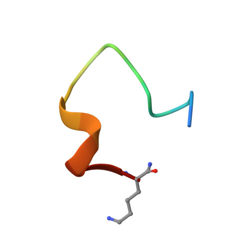
    Conformational changes of Aβ peptide result in its transformation from native monomeric state to the toxic soluble dimers, oligomers and insoluble aggregates that are hallmarks of Alzheimer's disease (AD). Interactions of zinc ions with Aβ are mediated by the N-terminal Aβ(1-16) domain and appear to play a key role in AD progression. There is a range of results indicating that these interactions trigger the Aβ plaque formation. We have determined structure and functional characteristics of the metal binding domains derived from several Aβ variants and found that their zinc-induced oligomerization is governed by conformational changes in the minimal zinc binding site 6HDSGYEVHH14. The residue H6 and segment 11EVHH14, which are part of this site are crucial for formation of the two zinc-mediated interaction interfaces in Aβ. These structural determinants can be considered as promising targets for rational design of the AD-modifying drugs aimed at blocking pathological Aβ aggregation.


  • Organizational Affiliation
    • Engelhardt Institute of Molecular Biology, Russian Academy of Sciences, 119991, Moscow, Russia.

Macromolecule Content 

  • Total Structure Weight: 4.07 kDa 
  • Atom Count: 287 
  • Modeled Residue Count: 36 
  • Deposited Residue Count: 36 
  • Unique protein chains: 1

Macromolecules

Find similar proteins by:  Sequence   |   3D Structure  
Entity ID: 1
MoleculeChains  Sequence LengthOrganismDetailsImage
Amyloid beta A4 protein
A, B
18Homo sapiensMutation(s): 3 
UniProt & NIH Common Fund Data Resources
Find proteins for P05067 (Homo sapiens)
Explore P05067 
Go to UniProtKB:  P05067
PHAROS:  P05067
GTEx:  ENSG00000142192 
Entity Groups
Sequence Clusters30% Identity50% Identity70% Identity90% Identity95% Identity100% Identity
UniProt GroupP05067
Sequence Annotations
Expand
Reference Sequence

Small Molecules

Ligands 1 Unique
IDChains Name / Formula / InChI Key2D Diagram3D Interactions
ZN

Query on ZN


Download:Ideal Coordinates CCD File
C [auth A]ZINC ION
Zn
PTFCDOFLOPIGGS-UHFFFAOYSA-N

Experimental Data & Validation

Experimental Data

  • Method: SOLUTION NMR
  • Conformers Calculated: 100 
  • Conformers Submitted: 20 
  • Selection Criteria: structures with the lowest energy 

Structure Validation

View Full Validation Report


Entry History 

Deposition Data

Revision History  (Full details and data files)

  • Version 1.0: 2014-11-12
    Type: Initial release
  • Version 1.1: 2016-03-02
    Changes: Database references
  • Version 1.2: 2020-01-01
    Changes: Data collection, Database references, Derived calculations
  • Version 1.3: 2024-10-30
    Changes: Data collection, Database references, Derived calculations, Structure summary