Probing the Dynamic Distribution of Bound States for Methylcytosine-binding Domains on DNA.
Cramer, J.M., Scarsdale, J.N., Walavalkar, N.M., Buchwald, W.A., Ginder, G.D., Williams, D.C.(2014) J Biological Chem 289: 1294-1302
- PubMed: 24307175 Search on PubMedSearch on PubMed Central
- DOI: https://doi.org/10.1074/jbc.M113.512236
- Primary Citation Related Structures:
2MB7 - PubMed Abstract:
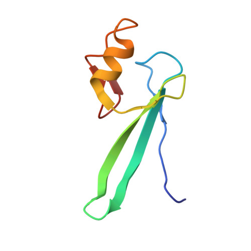
Although highly homologous to other methylcytosine-binding domain (MBD) proteins, MBD3 does not selectively bind methylated DNA, and thus the functional role of MBD3 remains in question. To explore the structural basis of its binding properties and potential function, we characterized the solution structure and binding distribution of the MBD3 MBD on hydroxymethylated, methylated, and unmethylated DNA. The overall fold of this domain is very similar to other MBDs, yet a key loop involved in DNA binding is more disordered than previously observed. Specific recognition of methylated DNA constrains the structure of this loop and results in large chemical shift changes in NMR spectra. Based on these spectral changes, we show that MBD3 preferentially localizes to methylated and, to a lesser degree, unmethylated cytosine-guanosine dinucleotides (CpGs), yet does not distinguish between hydroxymethylated and unmethylated sites. Measuring residual dipolar couplings for the different bound states clearly shows that the MBD3 structure does not change between methylation-specific and nonspecific binding modes. Furthermore, residual dipolar couplings measured for MBD3 bound to methylated DNA can be described by a linear combination of those for the methylation and nonspecific binding modes, confirming the preferential localization to methylated sites. The highly homologous MBD2 protein shows similar but much stronger localization to methylated as well as unmethylated CpGs. Together, these data establish the structural basis for the relative distribution of MBD2 and MBD3 on genomic DNA and their observed occupancy at active and inactive CpG-rich promoters.
- From the Department of Biochemistry and Molecular Biology.
Organizational Affiliation: