2LJ5 | pdb_00002lj5

Description of the Structural fluctuations of proteins from structure-based calculations of Residual dipolar couplings


Experimental Data Snapshot

  • Method: SOLUTION NMR
  • Conformers Calculated: 321 
  • Conformers Submitted: 301 
  • Selection Criteria: structures with acceptable covalent geometry 

wwPDB Validation 3D Report Full Report

Validation slider image for 2LJ5

This is version 1.2 of the entry. See complete history

Literature

Determination of structural fluctuations of proteins from structure-based calculations of residual dipolar couplings.

Montalvao, R.W.De Simone, A.Vendruscolo, M.

(2012) J Biomol NMR 53: 281-292

  • DOI: https://doi.org/10.1007/s10858-012-9644-3
  • Primary Citation Related Structures: 
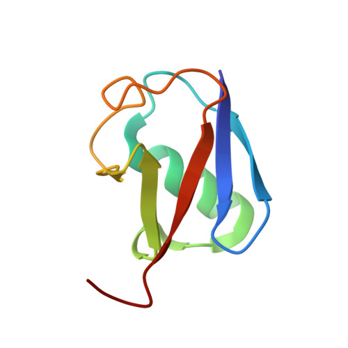
    2LJ5

  • PubMed Abstract: 

    Residual dipolar couplings (RDCs) have the potential of providing detailed information about the conformational fluctuations of proteins. It is very challenging, however, to extract such information because of the complex relationship between RDCs and protein structures. A promising approach to decode this relationship involves structure-based calculations of the alignment tensors of protein conformations. By implementing this strategy to generate structural restraints in molecular dynamics simulations we show that it is possible to extract effectively the information provided by RDCs about the conformational fluctuations in the native states of proteins. The approach that we present can be used in a wide range of alignment media, including Pf1, charged bicelles and gels. The accuracy of the method is demonstrated by the analysis of the Q factors for RDCs not used as restraints in the calculations, which are significantly lower than those corresponding to existing high-resolution structures and structural ensembles, hence showing that we capture effectively the contributions to RDCs from conformational fluctuations.


  • Organizational Affiliation
    • Department of Chemistry, University of Cambridge, Lensfield Road, Cambridge, CB2 1EW, UK.

Macromolecule Content 

  • Total Structure Weight: 8.58 kDa 
  • Atom Count: 602 
  • Modeled Residue Count: 76 
  • Deposited Residue Count: 76 
  • Unique protein chains: 1

Macromolecules

Find similar proteins by:|  3D Structure
Entity ID: 1
MoleculeChains  Sequence LengthOrganismDetailsImage
Ubiquitin76Homo sapiensMutation(s): 0 
Gene Names: UBA52UBCEP2RPS27AUBA80UBCEP1UBBUBC
UniProt & NIH Common Fund Data Resources
Find proteins for P62987 (Homo sapiens)
Explore P62987 
Go to UniProtKB:  P62987
PHAROS:  P62987
GTEx:  ENSG00000221983 
Entity Groups
Sequence Clusters30% Identity50% Identity70% Identity90% Identity95% Identity100% Identity
UniProt GroupP62987
Sequence Annotations
Expand
Reference Sequence

Experimental Data & Validation

Experimental Data

  • Method: SOLUTION NMR
  • Conformers Calculated: 321 
  • Conformers Submitted: 301 
  • Selection Criteria: structures with acceptable covalent geometry 

Structure Validation

View Full Validation Report


Entry History 

Deposition Data

Revision History  (Full details and data files)

  • Version 1.0: 2012-07-18
    Type: Initial release
  • Version 1.1: 2012-08-08
    Changes: Database references
  • Version 1.2: 2024-05-01
    Changes: Data collection, Database references