2KWV

Solution Structure of UBM1 of murine Polymerase iota in Complex with Ubiquitin


Experimental Data Snapshot

  • Method: SOLUTION NMR
  • Conformers Calculated: 200 
  • Conformers Submitted: 20 
  • Selection Criteria: structures with the lowest energy 

wwPDB Validation   3D Report Full Report


This is version 1.3 of the entry. See complete history


Literature

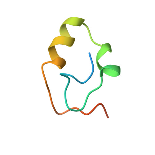
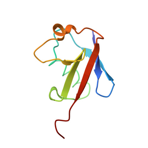
Structural analysis of the conserved ubiquitin-binding motifs (UBMs) of the translesion polymerase iota in complex with ubiquitin.

Burschowsky, D.Rudolf, F.Rabut, G.Herrmann, T.Matthias, P.Wider, G.

(2011) J Biol Chem 286: 1364-1373

  • DOI: https://doi.org/10.1074/jbc.M110.135038
  • Primary Citation of Related Structures:  
    2KWU, 2KWV

  • PubMed Abstract: 

    Ubiquitin-binding domains (UBDs) provide specificity to the ubiquitin system, which is also involved in translesion synthesis (TLS) in eukaryotic cells. Upon DNA damage, the UBDs (UBM domains) of polymerase iota (Pol ι) interact with ubiquitinated proliferating cell nuclear antigen to regulate the interchange between processive DNA polymerases and TLS. We report a biophysical analysis and solution structures of the two conserved UBM domains located in the C-terminal tail of murine Pol ι in complex with ubiquitin. The 35-amino acid core folds into a helix-turn-helix motif, which belongs to a novel domain fold. Similar to other UBDs, UBMs bind to ubiquitin on the hydrophobic surface delineated by Leu-8, Ile-44, and Val-70, however, slightly shifted toward the C terminus. In addition, UBMs also use electrostatic interactions to stabilize binding. NMR and fluorescence spectroscopy measurements revealed that UBMs bind monoubiquitin, and Lys-63- but not Lys-48-linked chains. Importantly, these biophysical data are supported by functional studies. Indeed, yeast cells expressing ubiquitin mutants specifically defective for UBM binding are viable but sensitive to DNA damaging conditions that require TLS for repair.


  • Organizational Affiliation

    Institute of Molecular Biology and Biophysics, ETH Zurich, 8093 Zurich, Switzerland.


Macromolecules
Find similar proteins by:  (by identity cutoff)  |  3D Structure
Entity ID: 1
MoleculeChains Sequence LengthOrganismDetailsImage
DNA polymerase iota48Mus musculusMutation(s): 1 
Gene Names: PoliRad30b
UniProt & NIH Common Fund Data Resources
Find proteins for Q6R3M4 (Mus musculus)
Explore Q6R3M4 
Go to UniProtKB:  Q6R3M4
IMPC:  MGI:1347081
Entity Groups  
Sequence Clusters30% Identity50% Identity70% Identity90% Identity95% Identity100% Identity
UniProt GroupQ6R3M4
Sequence Annotations
Expand
  • Reference Sequence
Find similar proteins by:  (by identity cutoff)  |  3D Structure
Entity ID: 2
MoleculeChains Sequence LengthOrganismDetailsImage
Ubiquitin76Homo sapiensMutation(s): 0 
Gene Names: RPS27AUBA80UBCEP1UBA52UBCEP2UBBUBC
UniProt & NIH Common Fund Data Resources
Find proteins for P0CG47 (Homo sapiens)
Explore P0CG47 
Go to UniProtKB:  P0CG47
PHAROS:  P0CG47
GTEx:  ENSG00000170315 
Entity Groups  
Sequence Clusters30% Identity50% Identity70% Identity90% Identity95% Identity100% Identity
UniProt GroupP0CG47
Sequence Annotations
Expand
  • Reference Sequence
Experimental Data & Validation

Experimental Data

  • Method: SOLUTION NMR
  • Conformers Calculated: 200 
  • Conformers Submitted: 20 
  • Selection Criteria: structures with the lowest energy 

Structure Validation

View Full Validation Report


Entry History 

Deposition Data

Revision History  (Full details and data files)

  • Version 1.0: 2010-10-06
    Type: Initial release
  • Version 1.1: 2011-07-13
    Changes: Version format compliance
  • Version 1.2: 2020-02-05
    Changes: Data collection, Database references, Other
  • Version 1.3: 2023-06-14
    Changes: Database references, Other