2K8B

Solution structure of PLAA family ubiquitin binding domain (PFUC) cis isomer in complex with ubiquitin


Experimental Data Snapshot

  • Method: SOLUTION NMR
  • Conformers Calculated: 1000 
  • Conformers Submitted: 10 
  • Selection Criteria: 10 structures for lowest energy 

wwPDB Validation   3D Report Full Report


This is version 1.2 of the entry. See complete history


Literature

Structural Basis for Ubiquitin Recognition by a Novel Domain from Human Phospholipase A2-activating Protein.

Fu, Q.S.Zhou, C.J.Gao, H.C.Jiang, Y.J.Zhou, Z.R.Hong, J.Yao, W.M.Song, A.X.Lin, D.H.Hu, H.Y.

(2009) J Biol Chem 284: 19043-19052

  • DOI: https://doi.org/10.1074/jbc.M109.009126
  • Primary Citation of Related Structures:  
    2K89, 2K8A, 2K8B, 2K8C

  • PubMed Abstract: 

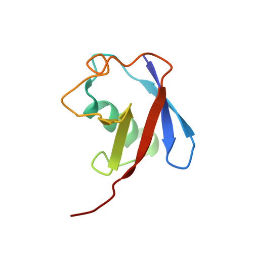
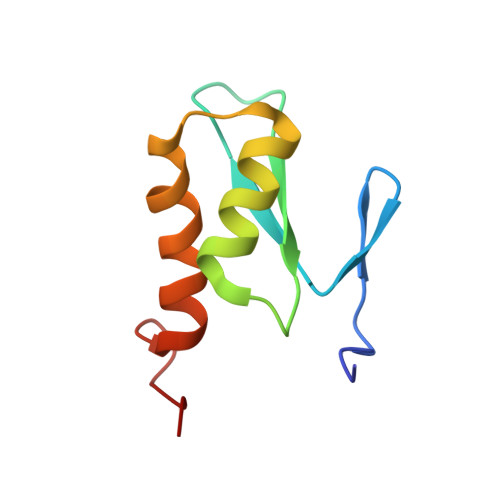
    Ubiquitin (Ub) is an essential modifier conserved in all eukaryotes from yeast to human. Phospholipase A(2)-activating protein (PLAA), a mammalian homolog of yeast DOA1/UFD3, has been proposed to be able to bind with Ub, which plays important roles in endoplasmic reticulum-associated degradation, vesicle formation, and DNA damage response. We have identified a core domain from the PLAA family ubiquitin-binding region of human PLAA (residues 386-465, namely PFUC) that can bind Ub and elucidated its solution structure and Ub-binding mode by NMR approaches. The PFUC domain possesses equal population of two conformers in solution by cis/trans-isomerization, whereas the two isomers exhibit almost equivalent Ub binding abilities. This domain structure takes a novel fold consisting of four beta-strands and two alpha-helices, and the Ub-binding site on PFUC locates in the surface of alpha2-helix, which is to some extent analogous to those of UBA, CUE, and UIM domains. This study provides structural basis and biochemical information for Ub recognition of the novel PFU domain from a PLAA family protein that may connect ubiquitination and degradation in endoplasmic reticulum-associated degradation.


  • Organizational Affiliation

    State Key Laboratory of Molecular Biology, Institute of Biochemistry and Cell Biology, Shanghai Institutes for Biological Sciences, Chinese Academy of Sciences, Shanghai 200031, China.


Macromolecules
Find similar proteins by:  (by identity cutoff)  |  3D Structure
Entity ID: 1
MoleculeChains Sequence LengthOrganismDetailsImage
Ubiquitin76Homo sapiensMutation(s): 0 
Gene Names: RPS27AUBA80UBCEP1UBA52UBCEP2UBBUBC
UniProt & NIH Common Fund Data Resources
Find proteins for P0CG48 (Homo sapiens)
Explore P0CG48 
Go to UniProtKB:  P0CG48
PHAROS:  P0CG48
GTEx:  ENSG00000150991 
Entity Groups  
Sequence Clusters30% Identity50% Identity70% Identity90% Identity95% Identity100% Identity
UniProt GroupP0CG48
Sequence Annotations
Expand
  • Reference Sequence
Find similar proteins by:  (by identity cutoff)  |  3D Structure
Entity ID: 2
MoleculeChains Sequence LengthOrganismDetailsImage
Phospholipase A-2-activating protein80Homo sapiensMutation(s): 0 
Gene Names: PLAAPLAP
UniProt & NIH Common Fund Data Resources
Find proteins for Q9Y263 (Homo sapiens)
Explore Q9Y263 
Go to UniProtKB:  Q9Y263
PHAROS:  Q9Y263
GTEx:  ENSG00000137055 
Entity Groups  
Sequence Clusters30% Identity50% Identity70% Identity90% Identity95% Identity100% Identity
UniProt GroupQ9Y263
Sequence Annotations
Expand
  • Reference Sequence
Experimental Data & Validation

Experimental Data

  • Method: SOLUTION NMR
  • Conformers Calculated: 1000 
  • Conformers Submitted: 10 
  • Selection Criteria: 10 structures for lowest energy 

Structure Validation

View Full Validation Report


Entry History 

Deposition Data

Revision History  (Full details and data files)

  • Version 1.0: 2009-05-05
    Type: Initial release
  • Version 1.1: 2011-07-13
    Changes: Version format compliance
  • Version 1.2: 2022-03-16
    Changes: Database references, Derived calculations