Protein Crystallization by Surface Entropy Reduction: Optimization of the Ser Strategy
Cooper, D.R., Boczek, T., Grelewska, K., Pinkowska, M., Sikorska, M., Zawadzki, M., Derewenda, Z.S.(2007) Acta Crystallogr D Biol Crystallogr 63: 636
- PubMed: 17452789 Search on PubMed
- DOI: https://doi.org/10.1107/S0907444907010931
- Primary Citation Related Structures:
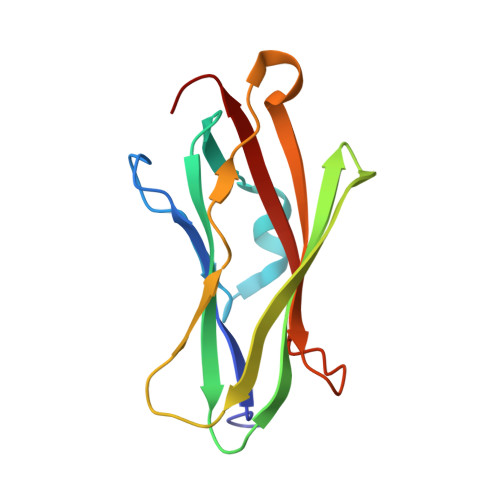
2BXW, 2JHS, 2JHT, 2JHU, 2JHV, 2JHW, 2JHX, 2JHY, 2JHZ, 2JI0 - PubMed Abstract:
A strategy of rationally engineering protein surfaces with the aim of obtaining mutants that are distinctly more susceptible to crystallization than the wild-type protein has previously been suggested. The strategy relies on replacing small clusters of two to three surface residues characterized by high conformational entropy with alanines. This surface entropy reduction (or SER) method has proven to be an effective salvage pathway for proteins that are difficult to crystallize. Here, a systematic comparison of the efficacy of using Ala, His, Ser, Thr and Tyr to replace high-entropy residues is reported. A total of 40 mutants were generated and screened using two different procedures. The results reaffirm that alanine is a particularly good choice for a replacement residue and identify tyrosines and threonines as additional candidates that have considerable potential to mediate crystal contacts. The propensity of these mutants to form crystals in alternative screens in which the normal crystallization reservoir solutions were replaced with 1.5 M NaCl was also examined. The results were impressive: more than half of the mutants yielded a larger number of crystals with salt as the reservoir solution. This method greatly increased the variety of conditions that yielded crystals. Taken together, these results suggest a powerful crystallization strategy that combines surface engineering with efficient screening using standard and alternate reservoir solutions.
- Department of Molecular Physiology and Biological Physics and Integrated Center for Structure-Function Innovation, University of Virginia, Charlottesville, Virginia 22908-0736, USA.
Organizational Affiliation: