2J5Y | pdb_00002j5y

Crystal structure of the GA module from F.magna


Experimental Data Snapshot

  • Method: X-RAY DIFFRACTION
  • Resolution: 1.40 Å
  • R-Value Free: 
    0.199 (Depositor), 0.199 (DCC) 
  • R-Value Work: 
    0.148 (Depositor), 0.146 (DCC) 
  • R-Value Observed: 
    0.151 (Depositor) 

Starting Model: experimental
View more details

wwPDB Validation 3D Report Full Report

Validation slider image for 2J5Y

This is version 1.3 of the entry. See complete history

Literature

Crystal Structure of a Bacterial Albumin-Binding Domain at 1.4A Resolution.

Cramer, J.F.Nordberg, P.A.Hajdu, J.Lejon, S.

(2007) FEBS Lett 581: 3178

  • DOI: https://doi.org/10.1016/j.febslet.2007.06.003
  • Primary Citation Related Structures: 
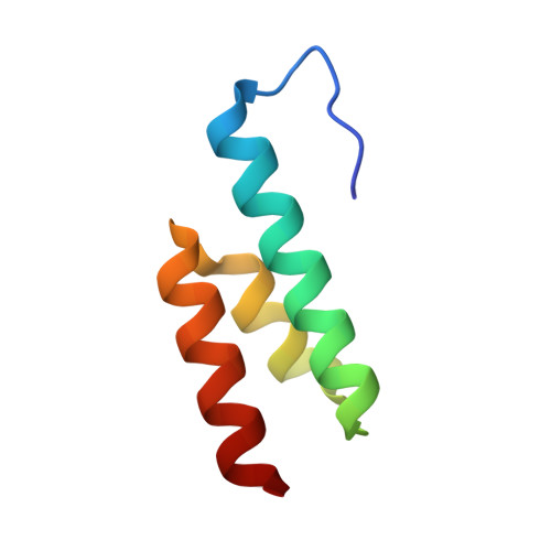
    2J5Y

  • PubMed Abstract: 

    The albumin-binding domain, or GA module, of the peptostreptococcal albumin-binding protein expressed in pathogenic strains of Finegoldia magna is believed to be responsible for the virulence and increased growth rate of these strains. Here we present the 1.4A crystal structure of this domain, and compare it with the crystal structure of the GA-albumin complex. An analysis of protein-protein interactions in the two crystals, and the presence of multimeric GA species in solution, indicate the GA module is "sticky", and is capable of forming contacts with a range of protein surfaces. This might lead to interactions with different host proteins.


  • Organizational Affiliation
    • Department of Cell and Molecular Biology, Uppsala University, Biomedical Centre, Uppsala, Sweden.

Macromolecule Content 

  • Total Structure Weight: 13.64 kDa 
  • Atom Count: 1,221 
  • Modeled Residue Count: 122 
  • Deposited Residue Count: 122 
  • Unique protein chains: 1

Macromolecules

Find similar proteins by:|  3D Structure
Entity ID: 1
MoleculeChains  Sequence LengthOrganismDetailsImage
PEPTOSTREPTOCOCCAL ALBUMIN-BINDING PROTEIN
A, B
61Finegoldia magnaMutation(s): 0 
UniProt
Find proteins for Q51911 (Finegoldia magna)
Explore Q51911 
Go to UniProtKB:  Q51911
Entity Groups
Sequence Clusters30% Identity50% Identity70% Identity90% Identity95% Identity100% Identity
UniProt GroupQ51911
Sequence Annotations
Expand
Reference Sequence

Experimental Data & Validation

Experimental Data

  • Method: X-RAY DIFFRACTION
  • Resolution: 1.40 Å
  • R-Value Free:  0.199 (Depositor), 0.199 (DCC) 
  • R-Value Work:  0.148 (Depositor), 0.146 (DCC) 
  • R-Value Observed: 0.151 (Depositor) 
Space Group: P 1 21 1
Unit Cell:
Length ( Å )Angle ( ˚ )
a = 25.784α = 90
b = 58.068β = 90.03
c = 40.57γ = 90
Software Package:
Software NamePurpose
REFMACrefinement
MOSFLMdata reduction
SCALAdata scaling
MOLREPphasing

Structure Validation

View Full Validation Report


Entry History 

Deposition Data

Revision History  (Full details and data files)

  • Version 1.0: 2007-06-26
    Type: Initial release
  • Version 1.1: 2011-05-08
    Changes: Version format compliance
  • Version 1.2: 2011-07-13
    Changes: Version format compliance
  • Version 1.3: 2023-12-13
    Changes: Data collection, Database references, Other, Refinement description