2J5M | pdb_00002j5m

Structure of Chloroperoxidase Compound 0


Experimental Data Snapshot

  • Method: X-RAY DIFFRACTION
  • Resolution: 1.75 Å
  • R-Value Free: 
    0.210 (Depositor), 0.209 (DCC) 
  • R-Value Work: 
    0.197 (Depositor), 0.197 (DCC) 
  • R-Value Observed: 
    0.198 (Depositor) 

Starting Model: experimental
View more details

wwPDB Validation 3D Report Full Report

Validation slider image for 2J5M

Ligand Structure Quality Assessment 


This is version 3.2 of the entry. See complete history

Literature

Structure and Quantum Chemical Characterization of Chloroperoxidase Compound 0, a Common Reaction Intermediate of Diverse Heme Enzymes.

Kuhnel, K.Derat, E.Terner, J.Shaik, S.Schlichting, I.

(2007) Proc Natl Acad Sci U S A 104: 99

  • DOI: https://doi.org/10.1073/pnas.0606285103
  • Primary Citation Related Structures: 
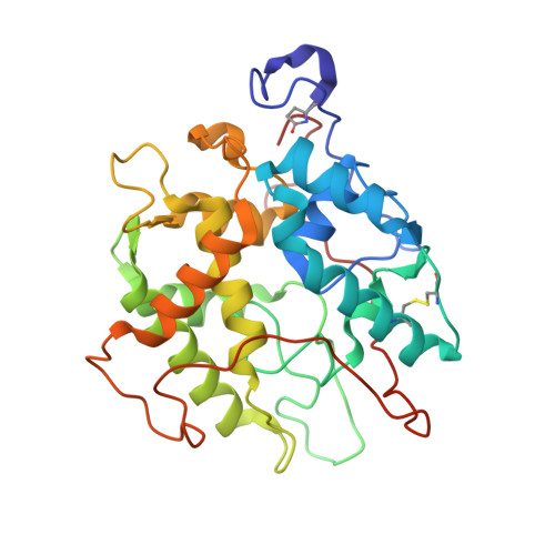
    2J5M

  • PubMed Abstract: 

    We have determined the crystal structure of the chloroperoxidase (CPO) hydroperoxo reaction intermediate (CPO compound 0) at 1.75-A resolution. The intermediate was generated through controlled photoreduction of the CPO oxygen complex during x-ray data collection, which was monitored by recording of the crystal absorption spectra. Initially, the peroxo-anion species was formed and then protonated to yield compound 0. Quantum chemical calculations indicate that the peroxo-anion species is not stable and collapses instantaneously to compound 0. Compound 0 is present in the ferric low-spin doublet ground state and is characterized by a long O O bond length of 1.5 A and a Fe O bond distance of 1.8 A, which is also observed in the crystal structure.


  • Organizational Affiliation
    • Department of Biomolecular Mechanisms, Max Planck Institute for Medical Research, Jahnstrasse 29, 69120 Heidelberg, Germany.

Macromolecule Content 

  • Total Structure Weight: 36.95 kDa 
  • Atom Count: 2,972 
  • Modeled Residue Count: 299 
  • Deposited Residue Count: 299 
  • Unique protein chains: 1

Macromolecules

Find similar proteins by:|  3D Structure
Entity ID: 1
MoleculeChains  Sequence LengthOrganismDetailsImage
CHLOROPEROXIDASE299Leptoxyphium fumagoMutation(s): 0 
EC: 1.11.1.10
UniProt
Find proteins for P04963 (Leptoxyphium fumago)
Explore P04963 
Go to UniProtKB:  P04963
Entity Groups
Sequence Clusters30% Identity50% Identity70% Identity90% Identity95% Identity100% Identity
UniProt GroupP04963
Glycosylation
Glycosylation Sites: 14
Sequence Annotations
Expand
Reference Sequence

Oligosaccharides

Help  
Entity ID: 2
MoleculeChains Length2D Diagram GlycosylationD Interactions
beta-D-mannopyranose-(1-4)-2-acetamido-2-deoxy-beta-D-glucopyranose-(1-4)-2-acetamido-2-deoxy-beta-D-glucopyranose
B
3N-Glycosylation
Glycosylation Resources
GlyTouCan: G15407YE
GlyCosmos: G15407YE
GlyGen: G15407YE
Entity ID: 3
MoleculeChains Length2D Diagram GlycosylationD Interactions
2-acetamido-2-deoxy-beta-D-glucopyranose-(1-4)-2-acetamido-2-deoxy-beta-D-glucopyranose
C
2N-Glycosylation
Glycosylation Resources
GlyTouCan: G42666HT
GlyCosmos: G42666HT
GlyGen: G42666HT
Entity ID: 4
MoleculeChains Length2D Diagram GlycosylationD Interactions
alpha-D-mannopyranose-(1-2)-alpha-D-mannopyranose
D
2O-Glycosylation
Glycosylation Resources
GlyTouCan: G53402KW
GlyCosmos: G53402KW
GlyGen: G53402KW

Small Molecules

Ligands 7 Unique
IDChains Name / Formula / InChI Key2D Diagram3D Interactions
HEM

Query on HEM


Download:Ideal Coordinates CCD File
F [auth A]PROTOPORPHYRIN IX CONTAINING FE
C34 H32 Fe N4 O4
KABFMIBPWCXCRK-RGGAHWMASA-L
NAG

Query on NAG


Download:Ideal Coordinates CCD File
H [auth A]2-acetamido-2-deoxy-beta-D-glucopyranose
C8 H15 N O6
OVRNDRQMDRJTHS-FMDGEEDCSA-N
MAN

Query on MAN


Download:Ideal Coordinates CCD File
I [auth A]
J [auth A]
K [auth A]
L [auth A]
M [auth A]
I [auth A],
J [auth A],
K [auth A],
L [auth A],
M [auth A],
N [auth A],
O [auth A],
P [auth A],
Q [auth A],
R [auth A]
alpha-D-mannopyranose
C6 H12 O6
WQZGKKKJIJFFOK-PQMKYFCFSA-N
EDO

Query on EDO


Download:Ideal Coordinates CCD File
S [auth A]1,2-ETHANEDIOL
C2 H6 O2
LYCAIKOWRPUZTN-UHFFFAOYSA-N
ACT

Query on ACT


Download:Ideal Coordinates CCD File
T [auth A]ACETATE ION
C2 H3 O2
QTBSBXVTEAMEQO-UHFFFAOYSA-M
MN

Query on MN


Download:Ideal Coordinates CCD File
E [auth A]MANGANESE (II) ION
Mn
WAEMQWOKJMHJLA-UHFFFAOYSA-N
PEO

Query on PEO


Download:Ideal Coordinates CCD File
G [auth A]HYDROGEN PEROXIDE
H2 O2
MHAJPDPJQMAIIY-UHFFFAOYSA-N
Modified Residues  1 Unique
IDChains TypeFormula2D DiagramParent
PCA
Query on PCA
A
L-PEPTIDE LINKINGC5 H7 N O3GLN

Biologically Interesting Molecules (External Reference) 

1 Unique

Experimental Data & Validation

Experimental Data

  • Method: X-RAY DIFFRACTION
  • Resolution: 1.75 Å
  • R-Value Free:  0.210 (Depositor), 0.209 (DCC) 
  • R-Value Work:  0.197 (Depositor), 0.197 (DCC) 
  • R-Value Observed: 0.198 (Depositor) 
Space Group: C 2 2 21
Unit Cell:
Length ( Å )Angle ( ˚ )
a = 59.01α = 90
b = 150.42β = 90
c = 99.59γ = 90
Software Package:
Software NamePurpose
REFMACrefinement
XDSdata reduction
XSCALEdata scaling
AMoREphasing

Structure Validation

View Full Validation Report


Ligand Structure Quality Assessment 


Entry History 

Deposition Data

Revision History  (Full details and data files)

  • Version 1.0: 2006-12-06
    Type: Initial release
  • Version 1.1: 2013-04-17
    Changes: Database references, Derived calculations, Non-polymer description, Other, Source and taxonomy, Structure summary, Version format compliance
  • Version 2.0: 2020-03-11
    Changes: Data collection, Derived calculations, Other, Polymer sequence
  • Version 3.0: 2020-07-29
    Type: Remediation
    Reason: Carbohydrate remediation
    Changes: Atomic model, Data collection, Derived calculations, Structure summary
  • Version 3.1: 2023-12-13
    Changes: Data collection, Database references, Refinement description, Structure summary
  • Version 3.2: 2024-11-13
    Changes: Structure summary