Sulfate Acts as Phosphate Analog on the Monomeric Catalytic Fragment of the Cpx-ATPase Copb from Sulfolobus Solfataricus
Luebben, M., Gueldenhaupt, J., Zoltner, M., Deigweiher, K., Haebel, P., Urbanke, C., Scheidig, A.J.(2007) J Mol Biol 369: 368
- PubMed: 17434529 Search on PubMed
- DOI: https://doi.org/10.1016/j.jmb.2007.03.029
- Primary Citation Related Structures:
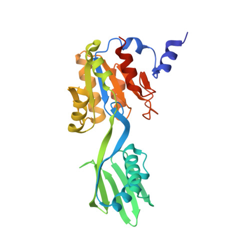
2IYE - PubMed Abstract:
The crystal structure of the catalytic fragment of a Sulfolobus solfataricus P-type ATPase, CopB-B, was determined with a 2.6 A resolution. CopB-B is the major soluble fragment of the archaeal CPx-ATPase CopB and is comprized of a nucleotide and a phosphorylation domain. In the crystalline state two molecules of CopB-B are in close contact to each other, although the presence of dimers in free solution could be ruled out by analytical ultracentrifugation. The overall architecture of CopB-B is similar to that of other P-type ATPases such as Ca-ATPase. Short peptide segments are linking the nucleotide binding to the phosphorylation domain. CopB-B exhibits 33% sequence identity (of 216 aligned residues) with the respective fragment of the Archaeoglobus fulgidus ATPase CopA. The CopB-B nucleotide-binding domain has the most primitive fold yet identified for this enzyme class. It is 24% identical to the nucleotide-binding domain of the disease-related Wilson ATPase ATP7B (80 structurally aligned residues). Structural superposition with Ca-ATPase suggests a putative nucleotide-binding site in CopB-B. The phosphorylation domain of CopB-B is structurally related to the corresponding part of Ca-ATPase in the anion-bound E2 state. In CopB-B crystals, a bound sulfate anion was identified at the phosphate-binding location. In solution state, the potential binding of CopB-B to phosphate was probed with (32)P(i). Bound phosphate could be readily displaced by orthovanadate at submillimolar concentration as well as by sulfate at millimolar concentration. It is possible therefore to assign the structure of the sulfate-bound phosphorylation domain of CopB-B to a state related to the E2.P(i) intermediate state of the catalytic cycle.
- Lehrstuhl für Biophysik, Ruhr-Universität Bochum, D-44780 Bochum, Germany. Mathias.Luebben@rub.de
Organizational Affiliation: