2IDO | pdb_00002ido

Structure of the E. coli Pol III epsilon-Hot proofreading complex


Experimental Data Snapshot

  • Method: X-RAY DIFFRACTION
  • Resolution: 2.10 Å
  • R-Value Free: 
    0.244 (Depositor), 0.247 (DCC) 
  • R-Value Work: 
    0.206 (Depositor) 
  • R-Value Observed: 
    0.209 (Depositor) 

Starting Model: experimental
View more details

wwPDB Validation 3D Report Full Report

Validation slider image for 2IDO

Ligand Structure Quality Assessment 


This is version 1.3 of the entry. See complete history

Literature

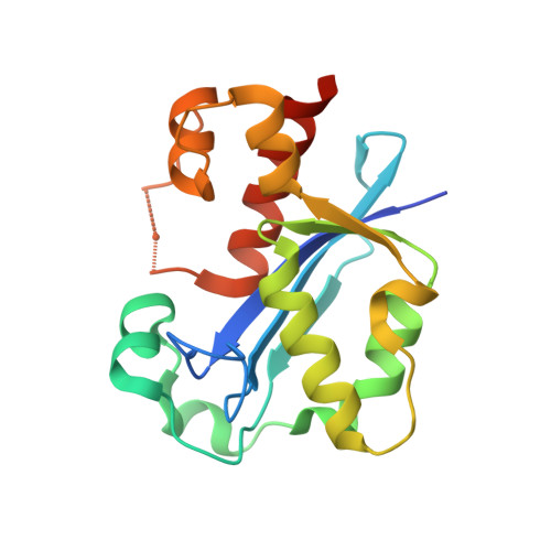
Structure of the Escherichia coli DNA polymerase III epsilon-HOT proofreading complex.

Kirby, T.W.Harvey, S.DeRose, E.F.Chalov, S.Chikova, A.K.Perrino, F.W.Schaaper, R.M.London, R.E.Pedersen, L.C.

(2006) J Biological Chem 281: 38466-38471

  • DOI: https://doi.org/10.1074/jbc.M606917200
  • Primary Citation Related Structures: 
    2IDO

  • PubMed Abstract: 

    The epsilon subunit of Escherichia coli DNA polymerase III possesses 3'-exonucleolytic proofreading activity. Within the Pol III core, epsilon is tightly bound between the alpha subunit (DNA polymerase) and subunit. Here, we present the crystal structure of epsilon in complex with HOT, the bacteriophage P1-encoded homolog of , at 2.1 A resolution. The epsilon-HOT interface is defined by two areas of contact: an interaction of the previously unstructured N terminus of HOT with an edge of the epsilon central beta-sheet as well as interactions between HOT and the catalytically important helix alpha1-loop-helix alpha2 motif of epsilon. This structure provides insight into how HOT and, by implication, may stabilize the epsilon subunit, thus promoting efficient proofreading during chromosomal replication.


  • Organizational Affiliation
    • Laboratory of Structural Biology, National Institute of Environmental Health Sciences/NIH, 111 TW Alexander Drive, Research Triangle Park, NC 27709, USA.

Macromolecule Content 

  • Total Structure Weight: 61.88 kDa 
  • Atom Count: 4,450 
  • Modeled Residue Count: 499 
  • Deposited Residue Count: 538 
  • Unique protein chains: 2

Macromolecules

Find similar proteins by:|  3D Structure
Entity ID: 1
MoleculeChains  Sequence LengthOrganismDetailsImage
DNA polymerase III epsilon subunit
A, C
186Escherichia coliMutation(s): 0 
Gene Names: dnaQmutD
EC: 2.7.7.7
UniProt
Find proteins for P03007 (Escherichia coli (strain K12))
Explore P03007 
Go to UniProtKB:  P03007
Entity Groups
Sequence Clusters30% Identity50% Identity70% Identity90% Identity95% Identity100% Identity
UniProt GroupP03007
Sequence Annotations
Expand
Reference Sequence
Find similar proteins by:|  3D Structure
Entity ID: 2
MoleculeChains  Sequence LengthOrganismDetailsImage
Hot protein
B, D
83Punavirus P1Mutation(s): 0 
Gene Names: hot
UniProt
Find proteins for Q71T70 (Escherichia phage P1)
Explore Q71T70 
Go to UniProtKB:  Q71T70
Entity Groups
Sequence Clusters30% Identity50% Identity70% Identity90% Identity95% Identity100% Identity
UniProt GroupQ71T70
Sequence Annotations
Expand
Reference Sequence

Small Molecules

Ligands 3 Unique
IDChains Name / Formula / InChI Key2D Diagram3D Interactions
TMP

Query on TMP


Download:Ideal Coordinates CCD File
G [auth A],
K [auth C]
THYMIDINE-5'-PHOSPHATE
C10 H15 N2 O8 P
GYOZYWVXFNDGLU-XLPZGREQSA-N
EDO

Query on EDO


Download:Ideal Coordinates CCD File
L [auth C]1,2-ETHANEDIOL
C2 H6 O2
LYCAIKOWRPUZTN-UHFFFAOYSA-N
MN

Query on MN


Download:Ideal Coordinates CCD File
E [auth A],
F [auth A],
H [auth C],
I [auth C],
J [auth C]
MANGANESE (II) ION
Mn
WAEMQWOKJMHJLA-UHFFFAOYSA-N

Experimental Data & Validation

Experimental Data

  • Method: X-RAY DIFFRACTION
  • Resolution: 2.10 Å
  • R-Value Free:  0.244 (Depositor), 0.247 (DCC) 
  • R-Value Work:  0.206 (Depositor) 
  • R-Value Observed: 0.209 (Depositor) 
Space Group: P 43 21 2
Unit Cell:
Length ( Å )Angle ( ˚ )
a = 77.289α = 90
b = 77.289β = 90
c = 212.978γ = 90
Software Package:
Software NamePurpose
CNSrefinement
StructureStudiodata collection
HKL-2000data reduction
HKL-2000data scaling
MOLREPphasing

Structure Validation

View Full Validation Report


Ligand Structure Quality Assessment 


Entry History 

Deposition Data

Revision History  (Full details and data files)

  • Version 1.0: 2006-11-14
    Type: Initial release
  • Version 1.1: 2008-05-01
    Changes: Version format compliance
  • Version 1.2: 2011-07-13
    Changes: Source and taxonomy, Version format compliance
  • Version 1.3: 2023-08-30
    Changes: Data collection, Database references, Derived calculations, Refinement description