2I25

Crystal structure analysis of the nurse shark New antigen Receptor PBLA8 variable domain in complex with lysozyme


Experimental Data Snapshot

  • Method: X-RAY DIFFRACTION
  • Resolution: 1.80 Å
  • R-Value Free: 0.211 
  • R-Value Work: 0.185 
  • R-Value Observed: 0.187 

wwPDB Validation   3D Report Full Report


This is version 1.2 of the entry. See complete history


Literature

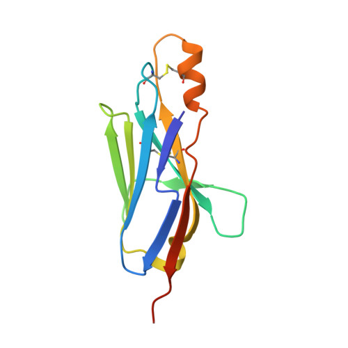
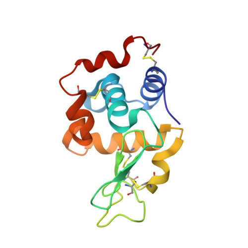
Maturation of Shark Single-domain (IgNAR) Antibodies: Evidence for Induced-fit Binding

Stanfield, R.L.Dooley, H.Verdino, P.Flajnik, M.F.Wilson, I.A.

(2007) J Mol Biol 367: 358-372

  • DOI: https://doi.org/10.1016/j.jmb.2006.12.045
  • Primary Citation of Related Structures:  
    2I24, 2I25, 2I26, 2I27

  • PubMed Abstract: 

    Sharks express an unusual heavy-chain isotype called IgNAR, whose variable regions bind antigen as independent soluble domains. To further probe affinity maturation of the IgNAR response, we structurally characterized the germline and somatically matured versions of a type II variable (V) region, both in the presence and absence of its antigen, hen egg-white lysozyme. Despite a disulfide bond linking complementarity determining regions (CDRs) 1 and 3, both germline and somatically matured V regions displayed significant structural changes in these CDRs upon complex formation with antigen. Somatic mutations in the IgNAR V region serve to increase the number of contacts with antigen, as reflected by a tenfold increase in affinity, and one of these mutations appears to stabilize the CDR3 region. In addition, a residue in the HV4 loop plays an important role in antibody-antigen interaction, consistent with the high rate of somatic mutations in this non-CDR loop.


  • Organizational Affiliation

    Department of Molecular Biology, The Scripps Research Institute, 10550 N. Torrey Pines Road, La Jolla, CA 92037, USA.


Macromolecules
Find similar proteins by:  (by identity cutoff)  |  3D Structure
Entity ID: 1
MoleculeChains Sequence LengthOrganismDetailsImage
New Antigen Receptor PBLA8A [auth N],
C [auth O]
121Ginglymostoma cirratumMutation(s): 0 
UniProt
Find proteins for Q8AXH5 (Ginglymostoma cirratum)
Explore Q8AXH5 
Go to UniProtKB:  Q8AXH5
Entity Groups  
Sequence Clusters30% Identity50% Identity70% Identity90% Identity95% Identity100% Identity
UniProt GroupQ8AXH5
Sequence Annotations
Expand
  • Reference Sequence
Find similar proteins by:  (by identity cutoff)  |  3D Structure
Entity ID: 2
MoleculeChains Sequence LengthOrganismDetailsImage
Lysozyme CB [auth L],
D [auth M]
129Gallus gallusMutation(s): 0 
EC: 3.2.1.17
UniProt
Find proteins for P00698 (Gallus gallus)
Explore P00698 
Go to UniProtKB:  P00698
Entity Groups  
Sequence Clusters30% Identity50% Identity70% Identity90% Identity95% Identity100% Identity
UniProt GroupP00698
Sequence Annotations
Expand
  • Reference Sequence
Experimental Data & Validation

Experimental Data

  • Method: X-RAY DIFFRACTION
  • Resolution: 1.80 Å
  • R-Value Free: 0.211 
  • R-Value Work: 0.185 
  • R-Value Observed: 0.187 
  • Space Group: P 21 21 21
Unit Cell:
Length ( Å )Angle ( ˚ )
a = 73.979α = 90
b = 82.719β = 90
c = 107.363γ = 90
Software Package:
Software NamePurpose
REFMACrefinement
Blu-Icedata collection
AMoREphasing

Structure Validation

View Full Validation Report


Entry History 

Deposition Data

Revision History  (Full details and data files)

  • Version 1.0: 2007-03-06
    Type: Initial release
  • Version 1.1: 2008-05-01
    Changes: Version format compliance
  • Version 1.2: 2011-07-13
    Changes: Advisory, Version format compliance