2HU1

Crystal structure Analysis of Hen Egg White Lyszoyme


Experimental Data Snapshot

  • Method: X-RAY DIFFRACTION
  • Resolution: 1.63 Å
  • R-Value Free: 0.220 
  • R-Value Work: 0.147 
  • R-Value Observed: 0.150 

wwPDB Validation   3D Report Full Report


This is version 1.3 of the entry. See complete history


Literature

Elucidation of the mechanism and end products of glutaraldehyde crosslinking reaction by X-ray structure analysis

Wine, Y.Cohen-Hadar, N.Freeman, A.Frolow, F.

(2007) Biotechnol Bioeng 98: 711-718

  • DOI: https://doi.org/10.1002/bit.21459
  • Primary Citation of Related Structures:  
    2HTX, 2HU1

  • PubMed Abstract: 

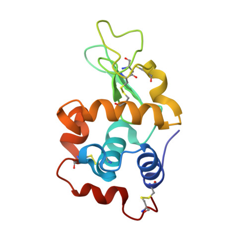
    Glutaraldehyde has been used for several decades as an effective crosslinking agent for many applications including sample fixation for microscopy, enzyme and cell immobilization, and stabilization of protein crystals. Despite of its common use as a crosslinking agent, the mechanism and chemistry involved in glutaraldehyde crosslinking reaction is not yet fully understood. Here we describe feasibility study and results obtained from a new approach to investigate the process of protein crystals stabilization by glutaraldehyde crosslinking. It involves exposure of a model protein crystal (Lysozyme) to glutaraldehyde in alkaline or acidic pH for different incubation periods and reaction arrest by medium exchange with crystallization medium to remove unbound glutaraldehyde. The crystals were subsequently incubated in diluted buffer affecting dissolution of un-crosslinked crystals. Samples from the resulting solution were subjected to protein composition analysis by gel electrophoresis and mass spectroscopy while crosslinked, dissolution resistant crystals were subjected to high resolution X-ray structural analysis. Data from gel electrophoresis indicated that the crosslinking process starts at specific preferable crosslinking site by lysozyme dimer formation, for both acidic and alkaline pH values. These dimer formations were followed by trimer and tetramer formations leading eventually to dissolution resistant crystals. The crosslinking initiation site and the end products obtained from glutaraldehyde crosslinking in both pH ranges resulted from reactions between lysine residues of neighboring protein molecules and the polymeric form of glutaraldehyde. Reaction rate was much faster at alkaline pH. Different reaction end products, indicating different reaction mechanisms, were identified for crosslinking taking place under alkaline or acidic conditions.


  • Organizational Affiliation

    Department of Molecular Microbiology and Biotechnology, Faculty of Life Sciences, Tel Aviv University, Tel Aviv, 69978, Israel.


Macromolecules
Find similar proteins by:  (by identity cutoff)  |  3D Structure
Entity ID: 1
MoleculeChains Sequence LengthOrganismDetailsImage
Lysozyme C129Gallus gallusMutation(s): 0 
EC: 3.2.1.17
UniProt
Find proteins for P00698 (Gallus gallus)
Explore P00698 
Go to UniProtKB:  P00698
Entity Groups  
Sequence Clusters30% Identity50% Identity70% Identity90% Identity95% Identity100% Identity
UniProt GroupP00698
Sequence Annotations
Expand
  • Reference Sequence
Small Molecules
Ligands 4 Unique
IDChains Name / Formula / InChI Key2D Diagram3D Interactions
220
Query on 220

Download Ideal Coordinates CCD File 
N [auth A]UNDECA-3,7-DIENE-1,3,7,11-TETRACARBALDEHYDE
C15 H20 O4
WRPMDTWVLJJHMV-UHFFFAOYSA-N
EDO
Query on EDO

Download Ideal Coordinates CCD File 
O [auth A],
P [auth A],
Q [auth A],
R [auth A],
S [auth A]
1,2-ETHANEDIOL
C2 H6 O2
LYCAIKOWRPUZTN-UHFFFAOYSA-N
CL
Query on CL

Download Ideal Coordinates CCD File 
J [auth A],
K [auth A],
L [auth A],
M [auth A]
CHLORIDE ION
Cl
VEXZGXHMUGYJMC-UHFFFAOYSA-M
NA
Query on NA

Download Ideal Coordinates CCD File 
B [auth A]
C [auth A]
D [auth A]
E [auth A]
F [auth A]
B [auth A],
C [auth A],
D [auth A],
E [auth A],
F [auth A],
G [auth A],
H [auth A],
I [auth A]
SODIUM ION
Na
FKNQFGJONOIPTF-UHFFFAOYSA-N
Experimental Data & Validation

Experimental Data

  • Method: X-RAY DIFFRACTION
  • Resolution: 1.63 Å
  • R-Value Free: 0.220 
  • R-Value Work: 0.147 
  • R-Value Observed: 0.150 
  • Space Group: P 43 21 2
Unit Cell:
Length ( Å )Angle ( ˚ )
a = 78.827α = 90
b = 78.827β = 90
c = 37.053γ = 90
Software Package:
Software NamePurpose
REFMACrefinement
HKL-2000data reduction
DENZOdata reduction
SCALEPACKdata scaling
REFMACphasing

Structure Validation

View Full Validation Report


Entry History 

Deposition Data

Revision History  (Full details and data files)

  • Version 1.0: 2007-05-22
    Type: Initial release
  • Version 1.1: 2007-11-20
    Changes: Version format compliance
  • Version 1.2: 2011-07-13
    Changes: Derived calculations, Version format compliance
  • Version 1.3: 2017-10-18
    Changes: Refinement description