2HJV | pdb_00002hjv

Structure of the second domain (residues 207-368) of the Bacillus subtilis YxiN protein


Experimental Data Snapshot

  • Method: X-RAY DIFFRACTION
  • Resolution: 1.95 Å
  • R-Value Free: 
    0.247 (Depositor), 0.246 (DCC) 
  • R-Value Work: 
    0.209 (Depositor), 0.209 (DCC) 
  • R-Value Observed: 
    0.209 (Depositor) 

Starting Model: experimental
View more details

wwPDB Validation 3D Report Full Report

Validation slider image for 2HJV

This is version 1.3 of the entry. See complete history

Literature

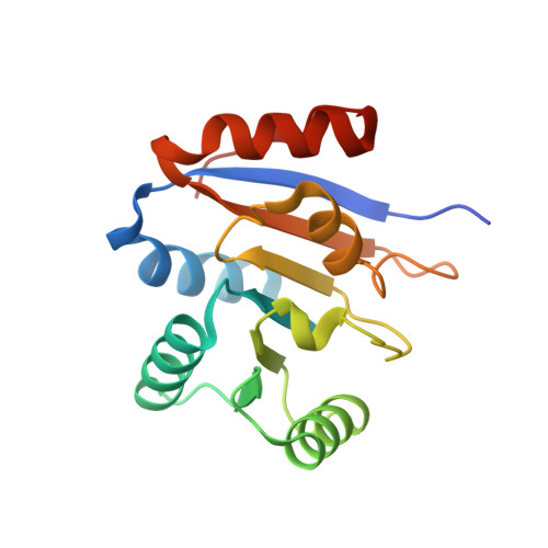
Structure of the second domain of the Bacillus subtilis DEAD-box RNA helicase YxiN.

Caruthers, J.M.Hu, Y.McKay, D.B.

(2006) Acta Crystallogr Sect F Struct Biol Cryst Commun 62: 1191-1195

  • DOI: https://doi.org/10.1107/S1744309106044642
  • Primary Citation Related Structures: 
    2HJV

  • PubMed Abstract: 

    The Bacillus subtilis RNA helicase YxiN is a modular three-domain protein. The first two domains form a conserved helicase core that couples an ATPase activity to an RNA duplex-destabilization activity, while the third domain recognizes a stem-loop of 23S ribosomal RNA with high affinity and specificity. The structure of the second domain, amino-acid residues 207-368, has been solved to 1.95 A resolution, revealing a parallel alphabeta-fold. The crystallographic asymmetric unit contains two protomers; superposition shows that they differ substantially in two segments of peptide that overlap the conserved helicase sequence motifs V and VI, while the remainder of the domain is isostructural. The conformational variability of these segments suggests that induced fit is intrinsic to the recognition of ligands (ATP and RNA) and the coupling of the ATPase activity to conformational changes.


  • Organizational Affiliation
    • Department of Structural Biology, Stanford University School of Medicine, Stanford, California 94305, USA.

Macromolecule Content 

  • Total Structure Weight: 37.35 kDa 
  • Atom Count: 2,696 
  • Modeled Residue Count: 313 
  • Deposited Residue Count: 326 
  • Unique protein chains: 1

Macromolecules

Find similar proteins by:|  3D Structure
Entity ID: 1
MoleculeChains  Sequence LengthOrganismDetailsImage
ATP-dependent RNA helicase dbpA
A, B
163Bacillus subtilisMutation(s): 1 
Gene Names: dbpA
EC: 3.6.1 (PDB Primary Data), 3.6.4.13 (UniProt)
UniProt
Find proteins for P42305 (Bacillus subtilis (strain 168))
Explore P42305 
Go to UniProtKB:  P42305
Entity Groups
Sequence Clusters30% Identity50% Identity70% Identity90% Identity95% Identity100% Identity
UniProt GroupP42305
Sequence Annotations
Expand
Reference Sequence

Experimental Data & Validation

Experimental Data

  • Method: X-RAY DIFFRACTION
  • Resolution: 1.95 Å
  • R-Value Free:  0.247 (Depositor), 0.246 (DCC) 
  • R-Value Work:  0.209 (Depositor), 0.209 (DCC) 
  • R-Value Observed: 0.209 (Depositor) 
Space Group: P 61
Unit Cell:
Length ( Å )Angle ( ˚ )
a = 105.2α = 90
b = 105.2β = 90
c = 66.03γ = 120
Software Package:
Software NamePurpose
CNSrefinement
HKL-2000data reduction
HKL-2000data scaling
CNSphasing

Structure Validation

View Full Validation Report


Entry History 

Deposition Data

Revision History  (Full details and data files)

  • Version 1.0: 2006-12-12
    Type: Initial release
  • Version 1.1: 2008-05-01
    Changes: Version format compliance
  • Version 1.2: 2011-07-13
    Changes: Version format compliance
  • Version 1.3: 2023-08-30
    Changes: Data collection, Database references, Refinement description