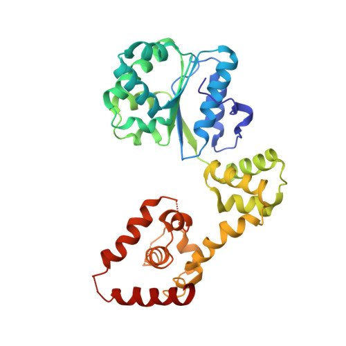
2HCB

Structure of AMPPCP-bound DnaA from Aquifex aeolicus


Experimental Data Snapshot

  • Method: X-RAY DIFFRACTION
  • Resolution: 3.51 Å
  • R-Value Free: 0.298 
  • R-Value Work: 0.266 
  • R-Value Observed: 0.267 

wwPDB Validation   3D Report Full Report


This is version 1.3 of the entry. See complete history


Literature

Structural basis for ATP-dependent DnaA assembly and replication-origin remodeling.

Erzberger, J.P.Mott, M.L.Berger, J.M.

(2006) Nat Struct Mol Biol 13: 676-683

  • DOI: https://doi.org/10.1038/nsmb1115
  • Primary Citation of Related Structures:  
    2HCB

  • PubMed Abstract: 

    In bacteria, the initiation of replication is controlled by DnaA, a member of the ATPases associated with various cellular activities (AAA+) protein superfamily. ATP binding allows DnaA to transition from a monomeric state into a large oligomeric complex that remodels replication origins, triggers duplex melting and facilitates replisome assembly. The crystal structure of AMP-PCP-bound DnaA reveals a right-handed superhelix defined by specific protein-ATP interactions. The observed quaternary structure of DnaA, along with topology footprint assays, indicates that a right-handed DNA wrap is formed around the initiation nucleoprotein complex. This model clarifies how DnaA engages and unwinds bacterial origins and suggests that additional, regulatory AAA+ proteins engage DnaA at filament ends. Eukaryotic and archaeal initiators also have the structural elements that promote open-helix formation, indicating that a spiral, open-ring AAA+ assembly forms the core element of initiators in all domains of life.


  • Organizational Affiliation

    Division of Biochemistry and Molecular Biology, Molecular and Cell Biology Department, 327B Hildebrand Hall #3206, University of California, Berkeley, California 94720, USA.


Macromolecules
Find similar proteins by:  (by identity cutoff)  |  3D Structure
Entity ID: 1
MoleculeChains Sequence LengthOrganismDetailsImage
Chromosomal replication initiator protein dnaA
A, B, C, D
323Aquifex aeolicusMutation(s): 0 
Gene Names: dnaA
UniProt
Find proteins for O66659 (Aquifex aeolicus (strain VF5))
Explore O66659 
Go to UniProtKB:  O66659
Entity Groups  
Sequence Clusters30% Identity50% Identity70% Identity90% Identity95% Identity100% Identity
UniProt GroupO66659
Sequence Annotations
Expand
  • Reference Sequence
Experimental Data & Validation

Experimental Data

  • Method: X-RAY DIFFRACTION
  • Resolution: 3.51 Å
  • R-Value Free: 0.298 
  • R-Value Work: 0.266 
  • R-Value Observed: 0.267 
  • Space Group: P 21 21 21
Unit Cell:
Length ( Å )Angle ( ˚ )
a = 99.076α = 90
b = 117.714β = 90
c = 200.994γ = 90
Software Package:
Software NamePurpose
REFMACrefinement
PDB_EXTRACTdata extraction
ADSCdata collection
HKL-2000data scaling
PHASERphasing

Structure Validation

View Full Validation Report


Entry History 

Deposition Data

Revision History  (Full details and data files)

  • Version 1.0: 2006-07-25
    Type: Initial release
  • Version 1.1: 2008-05-01
    Changes: Version format compliance
  • Version 1.2: 2011-07-13
    Changes: Advisory, Version format compliance
  • Version 1.3: 2023-08-30
    Changes: Data collection, Database references, Derived calculations, Refinement description