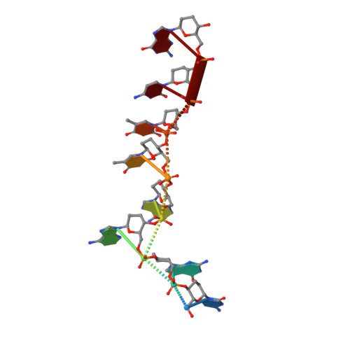
2H9S | pdb_00002h9s

Crystal Structure of Homo-DNA and Nature's Choice of Pentose over Hexose in the Genetic System

  • Classification: DNA
  • Mutation(s): No 

  • Deposited: 2006-06-11 Released: 2006-12-12 
  • Deposition Author(s): Egli, M., Pallan, P.S.

Experimental Data Snapshot

  • Method: X-RAY DIFFRACTION
  • Resolution: 1.75 Å
  • R-Value Free: 
    0.280 (Depositor) 
  • R-Value Work: 
    0.240 (Depositor), 0.240 (DCC) 
  • R-Value Observed: 
    0.240 (Depositor) 

wwPDB Validation 3D Report Full Report

Validation slider image for 2H9S

This is version 1.4 of the entry. See complete history. 

Literature

Crystal structure of homo-DNA and nature's choice of pentose over hexose in the genetic system.

Egli, M., Pallan, P.S., Pattanayek, R., Wilds, C.J., Lubini, P., Minasov, G., Dobler, M., Leumann, C.J., Eschenmoser, A.

(2006) J Am Chem Soc 128: 10847-10856

  • DOI: https://doi.org/10.1021/ja062548x
  • Primary Citation Related Structures: 
    2H9S

  • PubMed Abstract: 

    An experimental rationalization of the structure type encountered in DNA and RNA by systematically investigating the chemical and physical properties of alternative nucleic acids has identified systems with a variety of sugar-phosphate backbones that are capable of Watson-Crick base pairing and in some cases cross-pairing with the natural nucleic acids. The earliest among the model systems tested to date, (4' --> 6')-linked oligo(2',3'-dideoxy-beta-d-glucopyranosyl)nucleotides or homo-DNA, shows stable self-pairing, but the pairing rules for the four natural bases are not the same as those in DNA. However, a complete interpretation and understanding of the properties of the hexapyranosyl (4' --> 6') family of nucleic acids has been impeded until now by the lack of detailed 3D-structural data. We have determined the crystal structure of a homo-DNA octamer. It reveals a weakly twisted right-handed duplex with a strong inclination between the hexose-phosphate backbones and base-pair axes, and highly irregular values for helical rise and twist at individual base steps. The structure allows a rationalization of the inability of allo-, altro-, and glucopyranosyl-based oligonucleotides to form stable pairing systems.


  • Organizational Affiliation: 
    • Department of Biochemistry, School of Medicine, Vanderbilt University, Nashville, Tennessee 37232, USA. martin.egli@vanderbilt.edu

Macromolecule Content 

  • Total Structure Weight: 5.1 kDa 
  • Atom Count: 437 
  • Modeled Residue Count: 16 
  • Deposited Residue Count: 16 
  • Unique nucleic acid chains: 1

Macromolecules

Find similar nucleic acids by:  Sequence
Entity ID: 1
MoleculeChains LengthOrganismImage
5'-D(*(XCT)P*(XGU)P*(XAD)P*(XAD)P*(XTH)P*(XTH)P*(XCT)P*(XGU))-3'
A, B
8N/A
Sequence Annotations
Expand
Reference Sequence

Small Molecules

Ligands 1 Unique
IDChains Name / Formula / InChI Key2D Diagram3D Interactions
MG

Query on MG


Download:Ideal Coordinates CCD File
C [auth A]MAGNESIUM ION
Mg
JLVVSXFLKOJNIY-UHFFFAOYSA-N

Experimental Data & Validation

Experimental Data

  • Method: X-RAY DIFFRACTION
  • Resolution: 1.75 Å
  • R-Value Free:  0.280 (Depositor) 
  • R-Value Work:  0.240 (Depositor), 0.240 (DCC) 
  • R-Value Observed: 0.240 (Depositor) 
Space Group: P 61 2 2
Unit Cell:
Length ( Å )Angle ( ˚ )
a = 38.94α = 90
b = 38.94β = 90
c = 133.85γ = 120
Software Package:
Software NamePurpose
REFMACrefinement
HKL-2000data reduction
CNSrefinement
DENZOdata reduction
HKL-2000data scaling
CNSphasing

Structure Validation

View Full Validation Report


Entry History 

Deposition Data

Revision History  (Full details and data files)

  • Version 1.0: 2006-12-12
    Type: Initial release
  • Version 1.1: 2008-05-01
    Changes: Version format compliance
  • Version 1.2: 2011-07-13
    Changes: Version format compliance
  • Version 1.3: 2019-07-24
    Changes: Advisory, Data collection, Derived calculations, Refinement description
  • Version 1.4: 2024-02-14
    Changes: Advisory, Data collection, Database references, Derived calculations