2H4N

H94N CARBONIC ANHYDRASE II COMPLEXED WITH ACETAZOLAMIDE


Experimental Data Snapshot

  • Method: X-RAY DIFFRACTION
  • Resolution: 1.90 Å
  • R-Value Free: 0.219 
  • R-Value Work: 0.177 
  • R-Value Observed: 0.177 

wwPDB Validation   3D Report Full Report


This is version 1.4 of the entry. See complete history


Literature

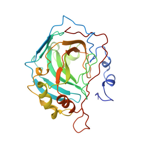
Histidine --> carboxamide ligand substitutions in the zinc binding site of carbonic anhydrase II alter metal coordination geometry but retain catalytic activity.

Lesburg, C.A.Huang, C.Christianson, D.W.Fierke, C.A.

(1997) Biochemistry 36: 15780-15791

  • DOI: https://doi.org/10.1021/bi971296x
  • Primary Citation of Related Structures:  
    1H4N, 1H9N, 1H9Q, 2H4N

  • PubMed Abstract: 

    The catalytic zinc ion of human carbonic anhydrase II (CAII) is coordinated by three histidine ligands (H94, H96, and H119) and a hydroxide ion with tetrahedral geometry. Structural and functional analysis of variants in which the zinc ligands H94 and H119 are substituted with asparagine and glutamine, and comparison with results obtained with aspartate and glutamate substitutions indicate that the neutral ligand field provided by the protein optimizes the electrostatic environment for the catalytic function of the metal ion, including stabilization of bound anions. This is demonstrated by catalytic activity measurements for ester hydrolysis and CO2 hydration, as well as sulfonamide inhibitor affinity assays. High-resolution X-ray crystal structure determinations of H94N, H119N, and H119Q CAIIs reveal that the engineered carboxamide side chains coordinate to zinc with optimal stereochemistry. However, zinc coordination geometry remains tetrahedral only in H119Q CAII. Metal geometry changes to trigonal bipyramidal in H119N CAII due to the addition of a second water molecule to the zinc coordination polyhedron and also in H94N CAII due to the displacement of zinc-bound hydroxide by the bidentate coordination of a Tris molecule. Possibly, the bulky histidine imidazole ligands of the native enzyme play a role in disfavoring trigonal bipyramidal coordination geometry for zinc. Protein-metal affinity is significantly compromised by all histidine --> carboxamide ligand substitutions. Diminished affinity may result from significant movements (up to 1 A) of the metal ion from its position in the wild-type enzyme, as well as the associated, minor conformational changes of metal ligands and their neighboring residues.


  • Organizational Affiliation

    Department of Chemistry, University of Pennsylvania, Philadelphia, Pennsylvania 19104-6323, USA.


Macromolecules
Find similar proteins by:  (by identity cutoff)  |  3D Structure
Entity ID: 1
MoleculeChains Sequence LengthOrganismDetailsImage
CARBONIC ANHYDRASE II259Homo sapiensMutation(s): 1 
Gene Names: CAII
EC: 4.2.1.1
UniProt & NIH Common Fund Data Resources
Find proteins for P00918 (Homo sapiens)
Explore P00918 
Go to UniProtKB:  P00918
PHAROS:  P00918
GTEx:  ENSG00000104267 
Entity Groups  
Sequence Clusters30% Identity50% Identity70% Identity90% Identity95% Identity100% Identity
UniProt GroupP00918
Sequence Annotations
Expand
  • Reference Sequence
Small Molecules
Ligands 2 Unique
IDChains Name / Formula / InChI Key2D Diagram3D Interactions
AZM
Query on AZM

Download Ideal Coordinates CCD File 
D [auth A]5-ACETAMIDO-1,3,4-THIADIAZOLE-2-SULFONAMIDE
C4 H6 N4 O3 S2
BZKPWHYZMXOIDC-UHFFFAOYSA-N
ZN
Query on ZN

Download Ideal Coordinates CCD File 
B [auth A],
C [auth A]
ZINC ION
Zn
PTFCDOFLOPIGGS-UHFFFAOYSA-N
Binding Affinity Annotations 
IDSourceBinding Affinity
AZM BindingDB:  2H4N Ki: min: 0.01, max: 104 (nM) from 25 assay(s)
Kd: min: 0.06, max: 46 (nM) from 12 assay(s)
IC50: min: 0.49, max: 4500 (nM) from 61 assay(s)
PDBBind:  2H4N Kd: 2 (nM) from 1 assay(s)
Binding MOAD:  2H4N Kd: 2.00e-3 (nM) from 1 assay(s)
Experimental Data & Validation

Experimental Data

  • Method: X-RAY DIFFRACTION
  • Resolution: 1.90 Å
  • R-Value Free: 0.219 
  • R-Value Work: 0.177 
  • R-Value Observed: 0.177 
  • Space Group: P 1 21 1
Unit Cell:
Length ( Å )Angle ( ˚ )
a = 42.7α = 90
b = 41.7β = 104.6
c = 73γ = 90
Software Package:
Software NamePurpose
X-PLORmodel building
X-PLORrefinement
MOSFLMdata reduction
CCP4data scaling
X-PLORphasing

Structure Validation

View Full Validation Report


Entry History 

Deposition Data

Revision History  (Full details and data files)

  • Version 1.0: 1997-09-17
    Type: Initial release
  • Version 1.1: 2008-03-24
    Changes: Version format compliance
  • Version 1.2: 2011-07-13
    Changes: Version format compliance
  • Version 1.3: 2021-11-03
    Changes: Database references, Derived calculations, Other
  • Version 1.4: 2023-08-09
    Changes: Refinement description